Randomized Controlled Trial
. 2012 Nov;117(5):981-95.
doi: 10.1097/ALN.0b013e31826be467.

Event-related functional magnetic resonance imaging of a low dose of dexmedetomidine that impairs long-term memory

Affiliations
Randomized Controlled Trial

Event-related functional magnetic resonance imaging of a low dose of dexmedetomidine that impairs long-term memory

Hiroki R Hayama et al. Anesthesiology. 2012 Nov.

Abstract

Background: Work suggests the amnesia from dexmedetomidine (an α2-adrenergic agonist) is caused by a failure of information to be encoded into long-term memory and that dexmedetomidine might differentially affect memory for emotionally arousing material. We investigated these issues in humans using event-related neuroimaging to reveal alterations in brain activity and subsequent memory effects associated with drug exposure.

Methods: Forty-eight healthy volunteers received a computer-controlled infusion of either placebo or low-dose dexmedetomidine (target = 0.15 ng/ml plasma) during neuroimaging while they viewed and rated 80 emotionally arousing (e.g., graphic war wound) and 80 nonarousing neutral (e.g., cup) pictures for emotional arousal content. Long-term picture memory was tested 4 days later without neuroimaging. Imaging data were analyzed for drug effects, emotional processing differences, and memory-related changes with statistical parametric mapping-8.

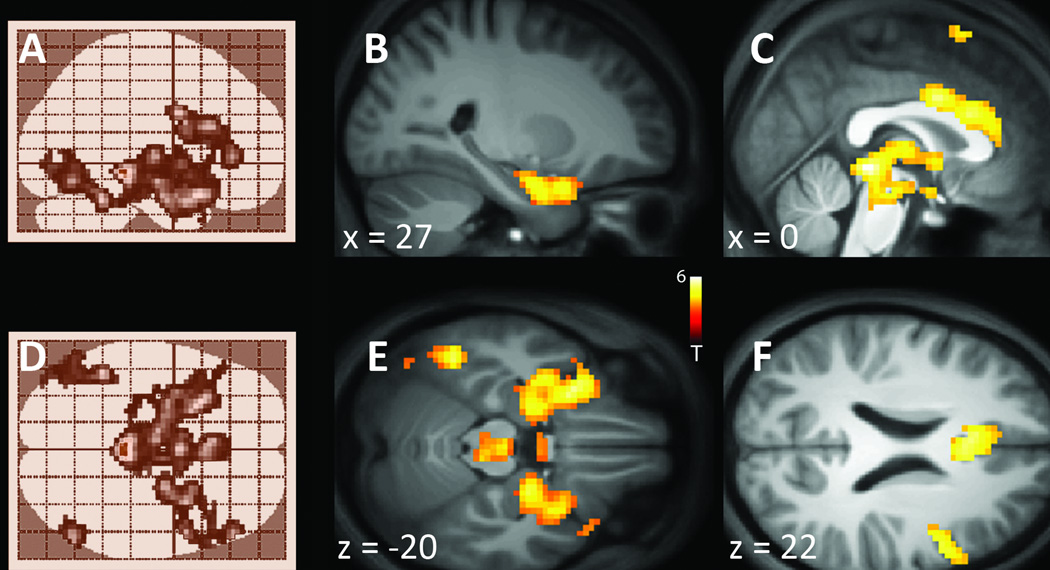
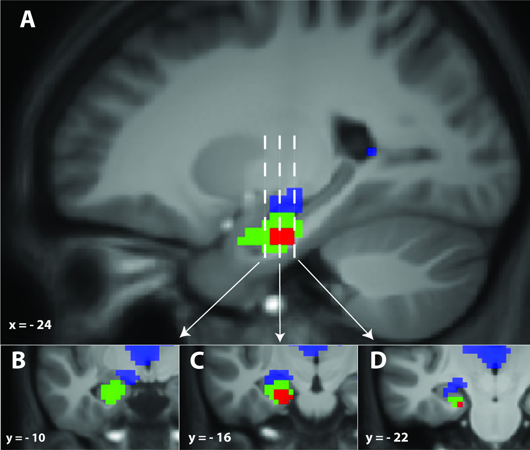
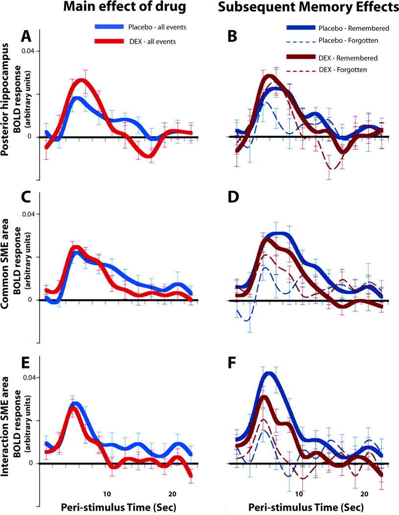
Results: Dexmedetomidine impaired overall (mean ± SEM) picture memory (placebo: 0.58 ± 0.03 vs. dexmedetomidine: 0.45 ± 0.03, P = 0.001), but did not differentially modulate memory as a function of item arousal. Arousing pictures were better remembered for both groups. Dexmedetomidine had regionally heterogeneous effects on brain activity, primarily decreasing it in the cortex and increasing it in thalamic and posterior hippocampal regions. Nevertheless, a single subsequent memory effect for item memory common to both groups was identified only in the left hippocampus/amygdala. Much of this effect was found to be larger for the placebo than dexmedetomidine group.

Conclusion: Dexmedetomidine impaired long-term picture memory, but did not disproportionately block memory for emotionally arousing items. The memory impairment on dexmedetomidine corresponds with a weakened hippocampal subsequent memory effect.


Figures

Fig. 1
Fig. 1
Brain regions where event-related functional magnetic resonance imaging signals are modulated by dexmedetomidine during the picture viewing and emotionality rating task. Across the whole brain the areas where the blood oxygen level dependent signal is attenuated by dexmedetomidine are illustrated in shades of blue and areas where the signal is enhanced by dexmedetomidine are shown in red. (A) Images are shown at a standard event-related functional magnetic resonance imaging (fMRI) significance threshold of p < 0.001, with a 20-voxel extent threshold. At this threshold, dexmedetomidine appears to enhance the signals present in the center parts of the brain including the thalamus and hippocampal areas. At the same time, dexmedetomidine appears to suppress activity across large portions of the cortex. (B) Images are shown at a liberal significance threshold of p < 0.05, uncorrected, for viewing purposes only. The activity is displayed on the normalized across-subjects mean anatomical image.
Fig. 2
Fig. 2
(A) Results from the main effect of drug analysis are displayed on an inflated rendering of cortical brain surface showing right and left hemispheres, respectively. Blue areas show where the blood oxygen level dependent signal response is suppressed on dexmedetomidine relative to placebo at a significance level of p < 0.001, with a voxel extent threshold of 20. The uncolored areas (light gray regions) do not show a significant effect of the drug. The peristimulus time course of activity in the measured signal is shown for two representative areas, one shows a main effect of the drug (the green dot area, within the blue color in the occipital lobe) and one shows no significant effect (the yellow dot area, within the gray area in the parietal lobe). (B) Time courses of the main drug effect for all events are plotted for activity from the representative areas shown as the green [x = 27, y = −91, z = 10] and yellow [x = −48, y = −73, z = 28] dots in panel ‘A’. (C) Time courses from the same areas shown in ‘B’, but now the events are parsed into those items that were remembered versus those that were forgotten. No significant subsequent memory effects are found in these cortical regions, regardless of whether or not a significant main effect of the drug was found (mean activity ± SEM). BOLD = blood oxygen level dependent; DEX = dexmedetomidine.
Fig. 3
Fig. 3
Results of the contrast examining where brain activity responses are significantly greater for emotionally arousing pictures compared with neutral non-arousing pictures. (A) The maximum intensity projection results are shown in a sagittal view that is displayed at a threshold of p < 0.001, with a 20-voxel extent. This displays the 3 dimensional results occurring across the whole brain as seen when looking through the brain from a side view. The outline of the brain is shown in light color. The significant regional effects are shown as the dark areas within this brain outline. (B) When the results shown in ‘A’ are projected onto a single parasagittal cross section of the group mean anatomical image it is evident that emotionally arousing pictures (compared to neutral pictures) significantly activate the amygdala. (C) When the results shown in ‘A’ are projected onto a midsagittal cross section, then activation of the anterior cingulate region just above the corpus collosum can be seen. Also evident in this slice is activation of thalamic and brain stem areas. (D) The same results as in ‘A’ are now shown in the axial plane, as if looking through the brain from the top down. (E) When the results shown in ‘D’ are projected onto the group mean anatomical image sliced in the axial plane the amygdala activation can clearly be seen as bilateral. Other brain stem areas of activation can also be seen. (F) The anterior cingulate activation is shown in the axial plane.
Figure 4
Figure 4
The only significant subsequent memory effect that was identified is located in the left hippocampus. (A) Sagittal view of the brain depicting the three different types of hippocampal effects found in comparing placebo with dexmedetomidine. The green colored areas represent those regions where blood oxygen level dependent activity was greater for items that were subsequently remembered versus items that were subsequently forgotten common to both the placebo subjects and the subjects on dexmedetomidine. The red shaded areas illustrate where there was a significant interaction, such that the placebo subjects had a larger subsequent memory effect than the dexmedetomidine subjects. Thus, this analysis identifies the red highlighted area as one neural correlate associated with the behavioral decrease in long-term memory performance that occurs on dexmedetomidine. The blue region is shown for comparison to reveal how nearby brain activity was greater for dexmedetomidine than placebo, even though this particular region did not show any subsequent memory effect. Three different close-up coronal views of the hippocampus are illustrated in the inserts; (B) where the y coordinate is −10, (C) at the y coordinate of −16, and (D) at the y coordinate of −22. Images are displayed on sections of the normalized across-subjects mean anatomical image and thresholded at a minimum of p < 0.001, with a 20-voxel extent threshold.
Figure 5
Figure 5
Representative time courses to all events (i.e., the blue areas of fig. 4) from a posterior region of the hippocampus which were greater for the dexmedetomidine than the placebo condition are shown in (A). For this same voxel (B), parsing the data into remembered and forgotten events revealed no significant subsequent memory effects. A representative blood oxygen level dependent response from the common subsequent memory effect area (i.e., the green areas of fig. 4) shows that the dexmedetomidine and placebo responses were essentially identical when collapsed across all event types (C). However, parsing the data into remembered and forgotten events (D) revealed that a significant subsequent memory effect was present for both placebo and dexmedetomidine. A representative signal from the interaction subsequent memory effect area (i.e., the red areas of fig. 4) shows that the dexmedetomidine and placebo responses did not significantly differ across all events (E). However, parsing the data into remembered and forgotten events (F) reveals a much larger subsequent memory effect for placebo than dexmedetomidine. BOLD = blood oxygen level dependent; DEX = dexmedetomidine; SME = subsequent memory effect.

Comment in

  • The problem with amnesia.
    Veselis RA. Veselis RA. Anesthesiology. 2012 Nov;117(5):940-1. doi: 10.1097/ALN.0b013e31826be6d6. Anesthesiology. 2012. PMID: 22929728 No abstract available.

References

    1. Eger EI, 2nd, Koblin DD, Harris RA, Kendig JJ, Pohorille A, Halsey MJ, Trudell JR. Hypothesis: Inhaled anesthetics produce immobility and amnesia by different mechanisms at different sites. Anesth Analg. 1997;84:915–918. - PubMed
    1. Alkire MT, Gorski LA. Relative amnesic potency of five inhalational anesthetics follows the Meyer-Overton rule. Anesthesiology. 2004;101:417–429. - PubMed
    1. Perouansky M, Rau V, Ford T, Oh SI, Perkins M, Eger EI, 2nd, Pearce RA. Slowing of the hippocampal theta rhythm correlates with anesthetic-induced amnesia. Anesthesiology. 2010;113:1299–1309. - PMC - PubMed
    1. Bekker AY, Kaufman B, Samir H, Doyle W. The use of dexmedetomidine infusion for awake craniotomy. Anesth Analg. 2001;92:1251–1253. - PubMed
    1. Grant SA, Breslin DS, MacLeod DB, Gleason D, Martin G. Dexmedetomidine infusion for sedation during fiberoptic intubation: A report of three cases. J Clin Anesth. 2004;16:124–126. - PubMed

Publication types

MeSH terms

Substances