

Synaptic integration in dendrites: exceptional need for speed
- PMID: 22930273
- PMCID: PMC3528977
- DOI: 10.1113/jphysiol.2012.229328
Synaptic integration in dendrites: exceptional need for speed
Abstract
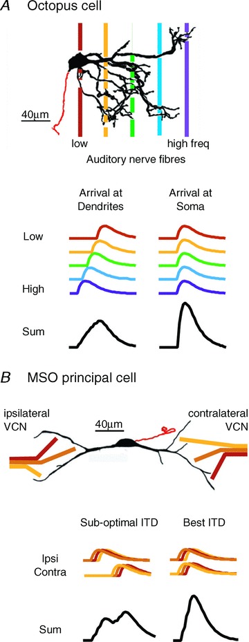
Some neurons in the mammalian auditory system are able to detect and report the coincident firing of inputs with remarkable temporal precision. A strong, low-voltage-activated potassium conductance (g(KL)) at the cell body and dendrites gives these neurons sensitivity to the rate of depolarization by EPSPs, allowing neurons to assess the coincidence of the rising slopes of unitary EPSPs. Two groups of neurons in the brain stem, octopus cells in the posteroventral cochlear nucleus and principal cells of the medial superior olive (MSO), extract acoustic information by assessing coincident firing of their inputs over a submillisecond timescale and convey that information at rates of up to 1000 spikes s(-1). Octopus cells detect the coincident activation of groups of auditory nerve fibres by broadband transient sounds, compensating for the travelling wave delay by dendritic filtering, while MSO neurons detect coincident activation of similarly tuned neurons from each of the two ears through separate dendritic tufts. Each makes use of filtering that is introduced by the spatial distribution of inputs on dendrites.
Figures
Similar articles
-
Generating synchrony from the asynchronous: compensation for cochlear traveling wave delays by the dendrites of individual brainstem neurons.J Neurosci. 2012 Jul 4;32(27):9301-11. doi: 10.1523/JNEUROSCI.0272-12.2012. J Neurosci. 2012. PMID: 22764237 Free PMC article.
-
Arrangement of Excitatory Synaptic Inputs on Dendrites of the Medial Superior Olive.J Neurosci. 2021 Jan 13;41(2):269-283. doi: 10.1523/JNEUROSCI.1055-20.2020. Epub 2020 Nov 18. J Neurosci. 2021. PMID: 33208467 Free PMC article.
-
Amplitude Normalization of Dendritic EPSPs at the Soma of Binaural Coincidence Detector Neurons of the Medial Superior Olive.J Neurosci. 2017 Mar 22;37(12):3138-3149. doi: 10.1523/JNEUROSCI.3110-16.2017. Epub 2017 Feb 17. J Neurosci. 2017. PMID: 28213442 Free PMC article.
-
Dendritic integration of excitatory synaptic input.Nat Rev Neurosci. 2000 Dec;1(3):181-90. doi: 10.1038/35044552. Nat Rev Neurosci. 2000. PMID: 11257906 Review.
-
The ion channels and synapses responsible for the physiological diversity of mammalian lower brainstem auditory neurons.Hear Res. 2019 May;376:33-46. doi: 10.1016/j.heares.2018.12.011. Epub 2018 Dec 26. Hear Res. 2019. PMID: 30606624 Review.
Cited by
-
A model of the medial superior olive explains spatiotemporal features of local field potentials.J Neurosci. 2014 Aug 27;34(35):11705-22. doi: 10.1523/JNEUROSCI.0175-14.2014. J Neurosci. 2014. PMID: 25164666 Free PMC article.
-
HCN channel-mediated neuromodulation can control action potential velocity and fidelity in central axons.Elife. 2019 Sep 9;8:e42766. doi: 10.7554/eLife.42766. Elife. 2019. PMID: 31496517 Free PMC article.
-
Physiology and anatomy of neurons in the medial superior olive of the mouse.J Neurophysiol. 2016 Dec 1;116(6):2676-2688. doi: 10.1152/jn.00523.2016. Epub 2016 Sep 21. J Neurophysiol. 2016. PMID: 27655966 Free PMC article.
-
Minimal conductance-based model of auditory coincidence detector neurons.PLoS One. 2015 Apr 6;10(4):e0122796. doi: 10.1371/journal.pone.0122796. eCollection 2015. PLoS One. 2015. PMID: 25844803 Free PMC article.
-
Cortical Specializations Underlying Fast Computations.Neuroscientist. 2016 Apr;22(2):145-64. doi: 10.1177/1073858415571539. Epub 2015 Feb 17. Neuroscientist. 2016. PMID: 25689988 Free PMC article. Review.
References
-
- Adams JC. Projections from octopus cells of the posteroventral cochlear nucleus to the ventral nucleus of the lateral lemniscus in cat and human. Auditory Neurosci. 1997;3:335–350.
-
- Agmon-Snir H, Carr CE, Rinzel J. The role of dendrites in auditory coincidence detection. Nature. 1998;393:268–272. - PubMed
-
- Bal R, Oertel D. Hyperpolarization-activated, mixed-cation current (Ih) in octopus cells of the mammalian cochlear nucleus. J Neurophysiol. 2000;84:806–817. - PubMed
-
- Bal R, Oertel D. Potassium currents in octopus cells of the mammalian cochlear nuclei. J Neurophysiol. 2001;86:2299–2311. - PubMed
-
- Brand A, Behrend O, Marquardt T, McAlpine D, Grothe B. Precise inhibition is essential for microsecond interaural time difference coding. Nature. 2002;417:543–547. - PubMed
Publication types
MeSH terms
Grants and funding
LinkOut - more resources
Full Text Sources