. 2012 Sep;140(3):325-39.
doi: 10.1085/jgp.201210851.

The contribution of hydrophobic residues in the pore-forming region of the ryanodine receptor channel to block by large tetraalkylammonium cations and Shaker B inactivation peptides

Affiliations

The contribution of hydrophobic residues in the pore-forming region of the ryanodine receptor channel to block by large tetraalkylammonium cations and Shaker B inactivation peptides

Sammy A Mason et al. J Gen Physiol. 2012 Sep.

Abstract

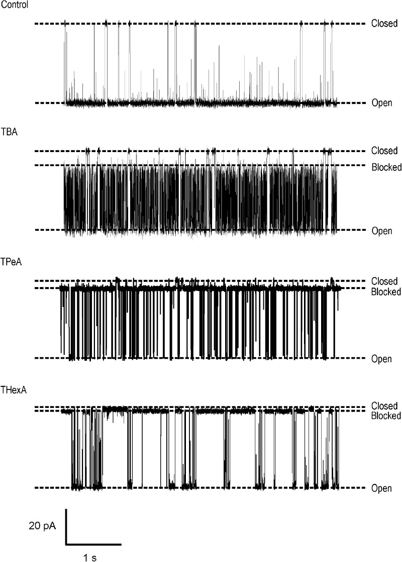
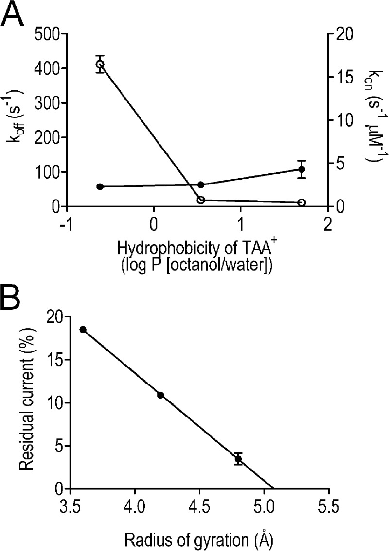
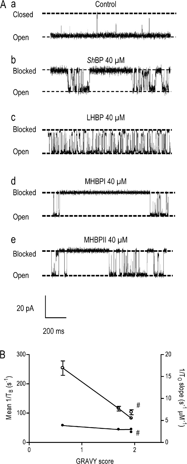
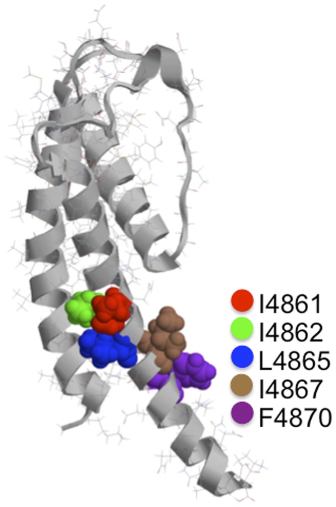
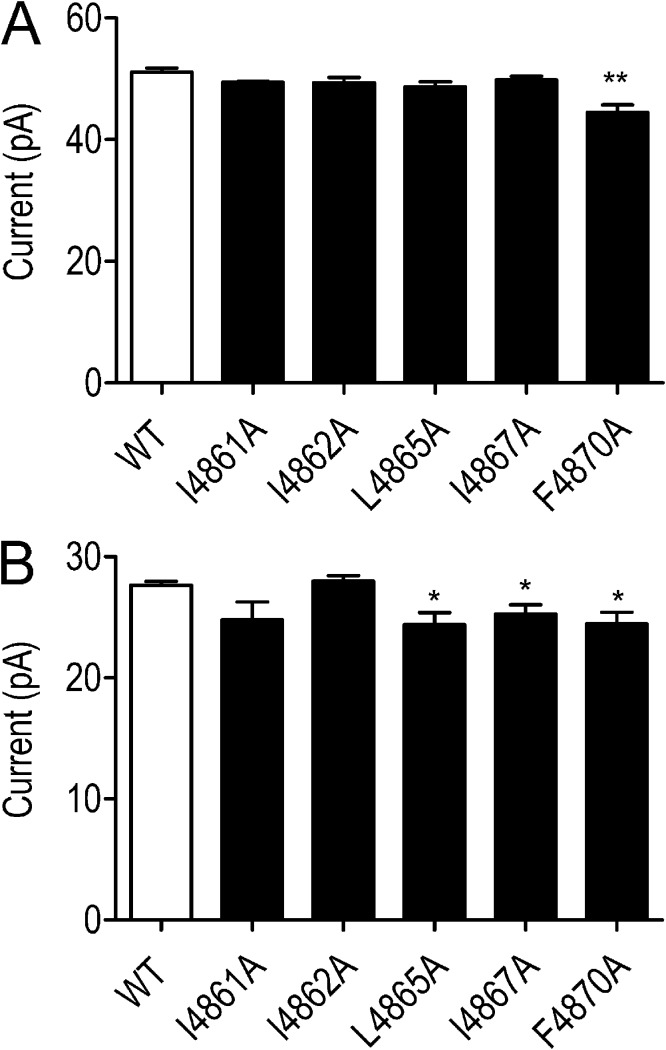
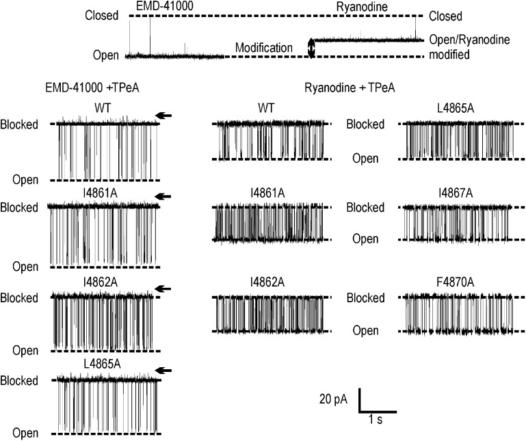
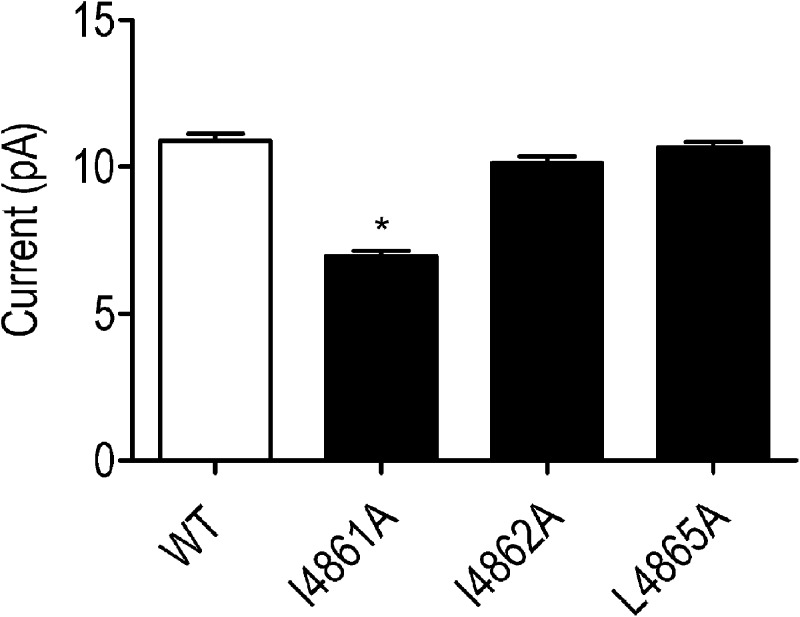
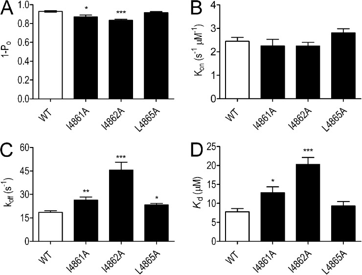
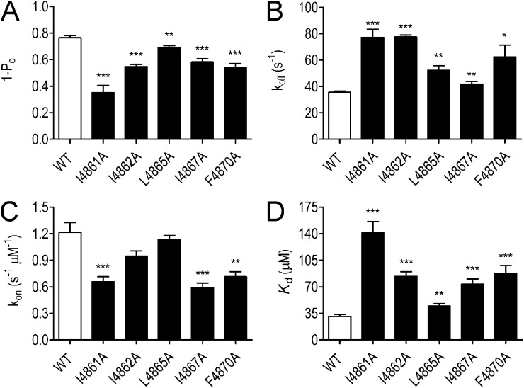
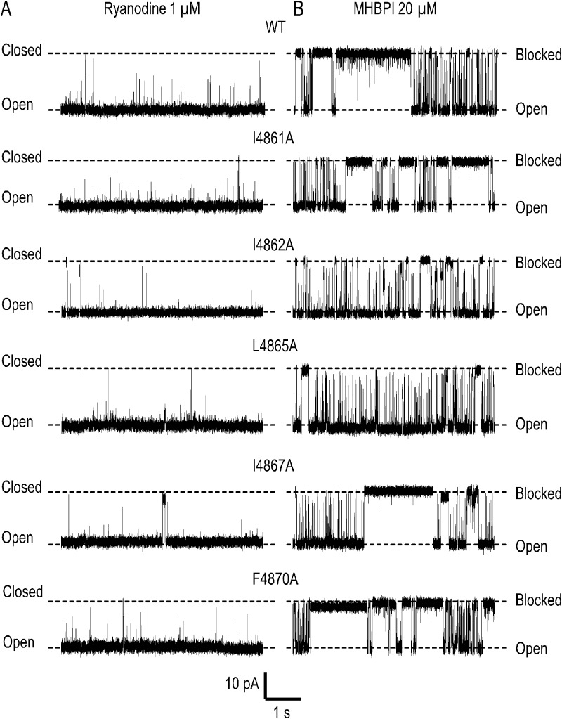
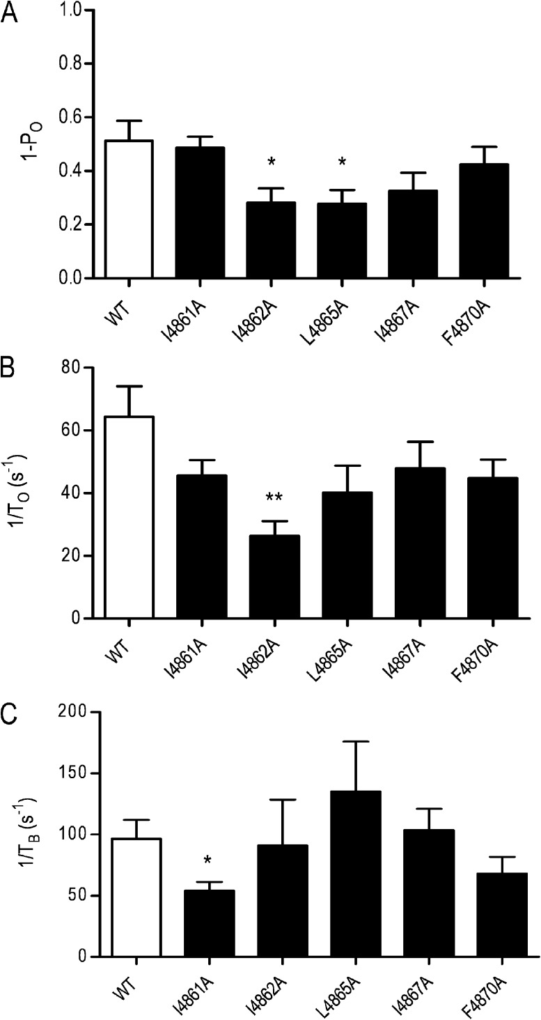
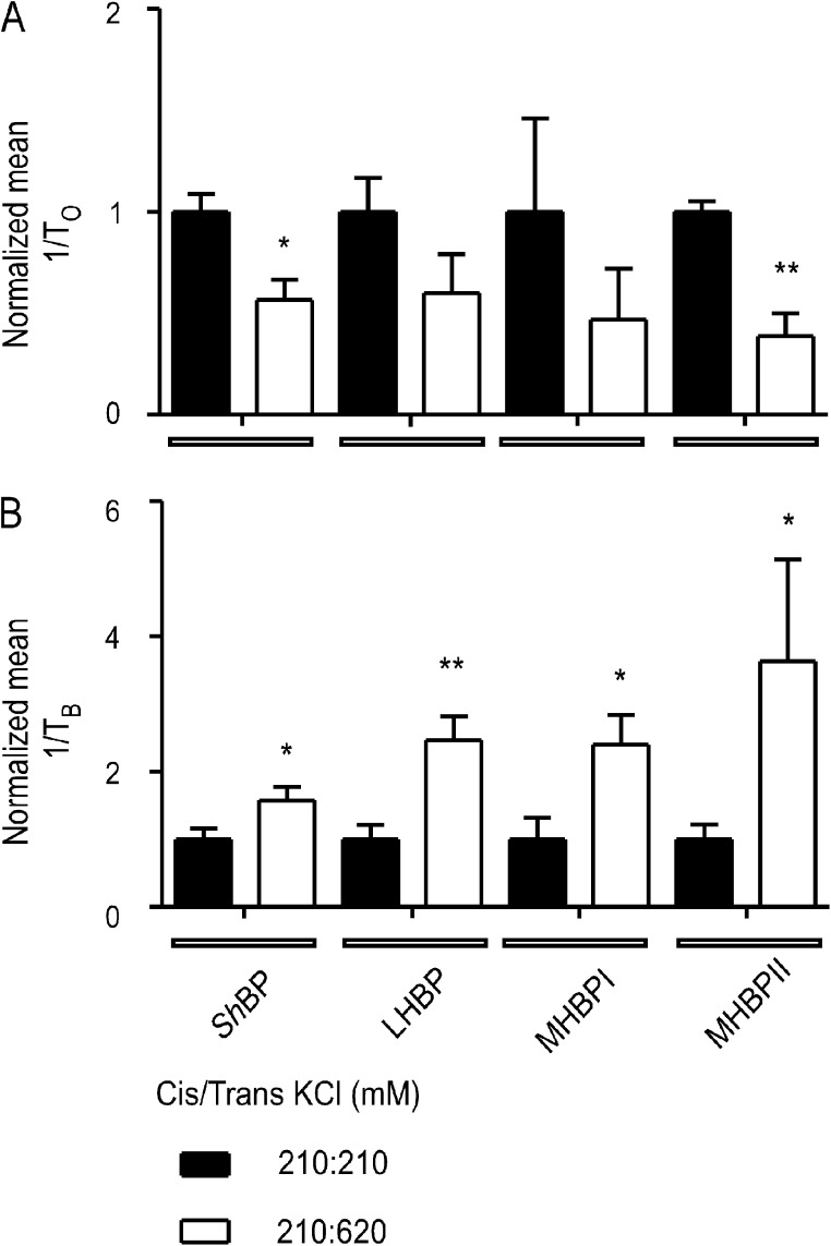
Although no high-resolution structural information is available for the ryanodine receptor (RyR) channel pore-forming region (PFR), molecular modeling has revealed broad structural similarities between this region and the equivalent region of K(+) channels. This study predicts that, as is the case in K(+) channels, RyR has a cytosolic vestibule lined with predominantly hydrophobic residues of transmembrane helices (TM10). In K(+) channels, this vestibule is the binding site for blocking tetraalkylammonium (TAA) cations and Shaker B inactivation peptides (ShBPs), which are stabilized by hydrophobic interactions involving specific residues of the lining helices. We have tested the hypothesis that the cytosolic vestibule of RyR fulfils a similar role and that TAAs and ShBPs are stabilized by hydrophobic interactions with residues of TM10. Both TAAs and ShBPs block RyR from the cytosolic side of the channel. By varying the composition of TAAs and ShBPs, we demonstrate that the affinity of both species is determined by their hydrophobicity, with variations reflecting alterations in the dissociation rate of the bound blockers. We investigated the role of TM10 residues of RyR by monitoring block by TAAs and ShBPs in channels in which the hydrophobicity of individual TM10 residues was lowered by alanine substitution. Although substitutions changed the kinetics of TAA interaction, they produced no significant changes in ShBP kinetics, indicating the absence of specific hydrophobic sites of interactions between RyR and these peptides. Our investigations (a) provide significant new information on both the mechanisms and structural components of the RyR PFR involved in block by TAAs and ShBPs, (b) highlight important differences in the mechanisms and structures determining TAA and ShBP block in RyR and K(+) channels, and (c) demonstrate that although the PFRs of these channels contain analogous structural components, significant differences in structure determine the distinct ion-handling properties of the two species of channel.


Figures

Figure 1.
Figure 1.
Large TAAs induce a characteristic partial block of K+ current in RyR2 channels. Single channel recordings of purified, EMD 41000–activated RyR2 channels at a holding potential of 60 mV with 610 mM K+ as the charge carrier. Addition of large TAAs (200 µM TBA, 100 µM TPeA, and 10 µM THexA) results in the occurrence of well-resolved blocking events.
Figure 2.
Figure 2.
Parameters of TAA block of RyR2 channels. (A) Rates of TAA association (closed circles, kon) and dissociation (open circles, koff) were determined at 60 mV and are plotted against cation hydrophobicity (monitored as the octanol/water partition coefficient). (B) The residual current after the interaction of 200 µM TBA, 100 µM TPeA, and 10 µM THexA was determined at 60 mV and expressed as a percentage of full open current and is plotted against the radius of gyration of the three TAAs (determined using SYBYL; Tripos). (A and B) Data are plotted as mean values (±SEM; n = 4 for TBA and THexA and n = 5 for TPeA).
Figure 3.
Figure 3.
Block of RyR2 channels by ShBPs. (A) Representative recordings of individual ryanodine-modified RyR2 channels in symmetrical 210 mM KCl (a) and after the addition of 40 µM ShBP (b), LHBP (c), MHBPI (d), and MHBPII (e) to the solution at the cytosolic (cis) side of the channel. In all cases, the holding potential was 50 mV. Closed, open, and blocked levels are indicated by dashed lines. (B) Rates of peptide association (1/To slope [per second/micromolar]; closed circles) and rates of peptide dissociation (mean 1/TB [per second]; open circles) are plotted against the GRAVY score of the hydrophobic portions of the four peptides: ShBP (1/To n = 3–11, 1/TB n = 3–13), LHBP (1/To n = 3–9, 1/TB n = 4–8), MHBPI (1/To n = 3–7, 1/TB n = 3–6), and MHBPII (1/To n = 4–10, 1/TB n = 4–9). Data are plotted as mean values (±SEM). As MHBPI and MHBPII have the same GRAVY score, MHBPII-related values are signified by “#.”
Figure 4.
Figure 4.
Location of hydrophobic residues in the proposed cytosolic cavity–lining helix (TM10) of RyR2. The figure shows one monomer of the Welch et al. (2004) model of the RyR2 PFR. The luminal entrance to the pore is at the top of the structure, and the cytosolic entrance is at the bottom. The hydrophobic residues of TM10 investigated in this study are highlighted, shown in space-fill, and identified in the key. Molecular graphics were rendered with Ras Top 2.0.2 software.
Figure 5.
Figure 5.
Current amplitudes in WT and residue-substituted channels. (A) Current amplitude of WT and TM10 alanine-substituted RyR2s at 60 mV after activation by 20 µM EMD 41000. (B) Equivalent current amplitudes after channel modification by 1 µM ryanodine. Data are plotted as mean values (±SEM; n = 4–7; *, P < 0.05; **, P < 0.01).
Figure 6.
Figure 6.
TPeA block of EMD 41000–activated and ryanodine-modified WT and substituted channels. The top panel shows representative traces of RyR2 in the absence of TPeA after addition of 20 µM EMD 41000 (left) and 1 µM ryanodine (right). The bottom left panel shows block of the WT and alanine-substituted EMD 41000–responsive channels by 100 µM TPeA at 60 mV. Closed levels are indicated by arrows. The bottom right panel shows representative traces of block of WT and substituted channels by 100 µM TPeA at 60 mV after modification by 1 µM ryanodine.
Figure 7.
Figure 7.
Residual current in WT and substituted EMD 41000–activated channels. Single channel current amplitudes of TPeA-blocked states monitored at 60 mV in the presence of 100 µM blocker. Data are plotted as mean values (±SEM; n = 4–7; *, P < 0.05).
Figure 8.
Figure 8.
The probability of block (1 − Po), rates of association/dissociation (kon/koff), and the affinity (Kd) of TPeA in EMD 41000–activated WT and substituted RyR2 channels. (A–D) Blocking parameters were determined at 60 mV in the presence of 100 µM TPeA. Data are plotted as mean values (±SEM; n = 5–7; *, P < 0.05; **, P < 0.01; ***, P < 0.001).
Figure 9.
Figure 9.
The probability of block (1 − Po), rates of association/dissociation (kon/koff), and the affinity (Kd) of TPeA in ryanodine-modified WT and substituted RyR2 channels. (A–D) Blocking parameters were determined at 60 mV in the presence of 100 µM TPeA. Data are plotted as mean values (±SEM; n = 5–8; *, P < 0.05; **, P < 0.01; ***, P < 0.001).
Figure 10.
Figure 10.
Effect of lowering the hydrophobicity of TM10 residues on the block of ryanodine-modified RyR2 channels by MHBPI. Representative single channel traces at a holding potential of 50 mV in symmetrical 210 mM KCl. MHBPI was applied to the cytosolic (cis) side of the RyR2 channels. Closed, open, and blocked levels are indicated by dashed lines. In each case, A shows representative activity of a ryanodine-modified channel before application of 20 µM MHBPI. B shows activity of the same channel after addition of the peptide.
Figure 11.
Figure 11.
Analysis of the parameters of MHBPI block after alanine substitution of TM10 residues. (A–C) Parameters of block were determined for WT and alanine-substituted ryanodine-modified channels induced by 20 µM MHBPI at a holding potential of 50 mV in symmetrical 210 mM KCl. Data are plotted as mean ± SEM for between five and eight channels. When significant differences occur between the WT parameter and that of the substituted channel, this is indicated by asterisks (*, P < 0.05; **, P < 0.01).
Figure 12.
Figure 12.
Destabilization of bound peptide analogues by increased luminal to cytosolic K+ flux through ryanodine-modified RyR2 channels. (A) Normalized values of the pooled rates of association (1/To) for 20 µM ShBP, 85 µM LHBP, 45 µM MHBPI, and 45 µM MHBPII monitored at 50 mV in either symmetrical 210 mM KCl or with 210 mM KCl in the cis chamber and 620 mM KCl in the trans chamber. (B) Normalized values of the pooled rates of dissociation (1/TB) under the same conditions. Data are plotted as mean ± SEM for between three and eight channels. Each individual value (for 210:210 and 210:620) was normalized to the mean value of the 210:210 data for each peptide. Where differences between values obtained in 210:210 and 210:620 reached statistical significance, these are indicated by asterisks (*, P < 0.05; **, P < 0.01).

Similar articles

Cited by

References

    1. Armstrong C.M. 1971. Interaction of tetraethylammonium ion derivatives with the potassium channels of giant axons. J. Gen. Physiol. 58:413–437 10.1085/jgp.58.4.413 - DOI - PMC - PubMed
    1. Bers D.M. 2002. Cardiac excitation-contraction coupling. Nature. 415:198–205 10.1038/415198a - DOI - PubMed
    1. Chen S.R.W., Li P., Zhao M.C., Li X.L., Zhang L. 2002. Role of the proposed pore-forming segment of the Ca2+ release channel (ryanodine receptor) in ryanodine interaction. Biophys. J. 82:2436–2447 10.1016/S0006-3495(02)75587-2 - DOI - PMC - PubMed
    1. Choi K.L., Mossman C., Aubé J., Yellen G. 1993. The internal quaternary ammonium receptor site of Shaker potassium channels. Neuron. 10:533–541 10.1016/0896-6273(93)90340-W - DOI - PubMed
    1. Cordero-Morales J.F., Cuello L.G., Zhao Y., Jogini V., Cortes D.M., Roux B., Perozo E. 2006. Molecular determinants of gating at the potassium-channel selectivity filter. Nat. Struct. Mol. Biol. 13:311–318 10.1038/nsmb1069 - DOI - PubMed

Publication types

MeSH terms

Substances