. 2012 Aug 29:12:378.
doi: 10.1186/1471-2407-12-378.

miRNA-21 is developmentally regulated in mouse brain and is co-expressed with SOX2 in glioma

Affiliations

miRNA-21 is developmentally regulated in mouse brain and is co-expressed with SOX2 in glioma

Jelena Põlajeva et al. BMC Cancer. .

Abstract

Background: MicroRNAs (miRNAs) and their role during tumor development have been studied in great detail during the last decade, albeit their expression pattern and regulation during normal development are however not so well established. Previous studies have shown that miRNAs are differentially expressed in solid human tumors. Platelet-derived growth factor (PDGF) signaling is known to be involved in normal development of the brain as well as in malignant primary brain tumors, gliomas, but the complete mechanism is still lacking. We decided to investigate the expression of the oncogenic miR-21 during normal mouse development and glioma, focusing on PDGF signaling as a potential regulator of miR-21.

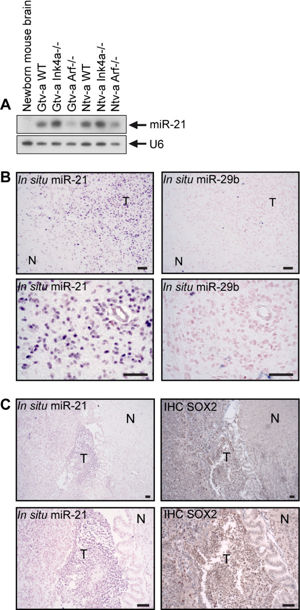
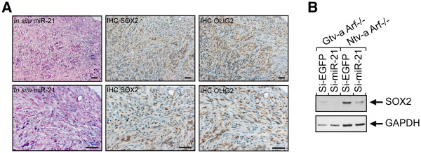
Methods: We generated mouse glioma using the RCAS/tv-a system for driving PDGF-BB expression in a cell-specific manner. Expression of miR-21 in mouse cell cultures and mouse brain were assessed using Northern blot analysis and in situ hybridization. Immunohistochemistry and Western blot analysis were used to investigate SOX2 expression. LNA-modified siRNA was used for irreversible depletion of miR-21. For inhibition of PDGF signaling Gleevec (imatinib mesylate), Rapamycin and U0126, as well as siRNA were used. Statistical significance was calculated using double-sided unpaired Student's t-test.

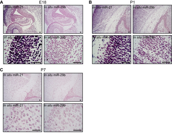
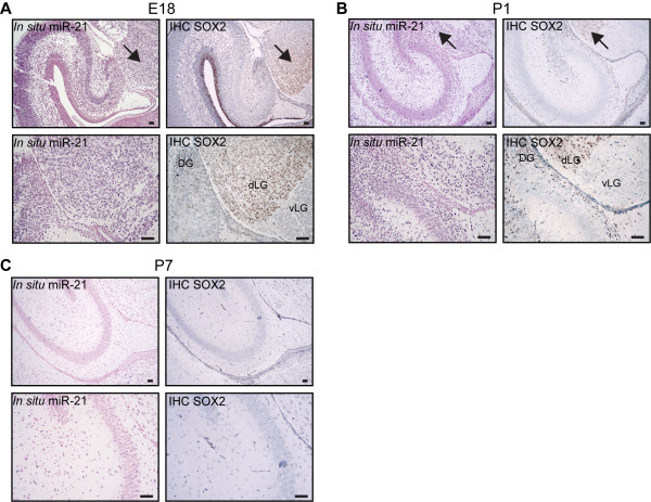
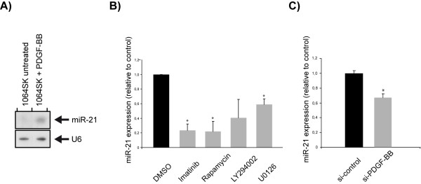
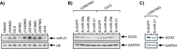
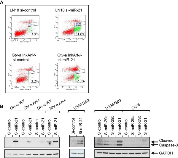
Results: We identified miR-21 to be highly expressed during embryonic and newborn brain development followed by a gradual decrease until undetectable at postnatal day 7 (P7), this pattern correlated with SOX2 expression. Furthermore, miR-21 and SOX2 showed up-regulation and overlapping expression pattern in RCAS/tv-a generated mouse brain tumor specimens. Upon irreversible depletion of miR-21 the expression of SOX2 was strongly diminished in both mouse primary glioma cultures and human glioma cell lines. Interestingly, in normal fibroblasts the expression of miR-21 was induced by PDGF-BB, and inhibition of PDGF signaling in mouse glioma primary cultures resulted in suppression of miR-21 suggesting that miR-21 is indeed regulated by PDGF signaling.

Conclusions: Our data show that miR-21 and SOX2 are tightly regulated already during embryogenesis and define a distinct population with putative tumor cell of origin characteristics. Furthermore, we believe that miR-21 is a mediator of PDGF-driven brain tumors, which suggests miR-21 as a promising target for treatment of glioma.


Figures

Figure 1
Figure 1
miR-21 is expressed during embryonic development.In situ hybridization with a digoxigenin-labeled probe, showing differential expression of miR-21 in normal E18 (A), newborn (P1) (B) and P7 (C) Gtv a wild type mouse brain. miR-21 could be seen in hippocampus as well as areas close to the ventricle. A digoxigenin miR-29b was used as an independent miRNA control. Scale bar is indicated in the figure and represents 50 μm.
Figure 2
Figure 2
Overlapping expression of SOX2 and miR-21. IHC and in situ hybridization showing the expression pattern of SOX2 and miR-21, respectively. miR-21 expression in Gtv a wild type mouse brain at different developmental stages E18 is shown in A), P1 in B) and P7 in C). Scale bar is indicated in the figure and represents 50 μm. Arrow indicates overlapping expression of SOX2 and miR-21. Abbreviations: dorsal lateral geniculate nucleus (dLG), ventral lateral geniculate nucleus (vLG), dentate gyrus (DG).
Figure 3
Figure 3
Increased levels of miR-21 in mouse glioma cell lines and glioma. A) Compared to normal mouse brain the different mouse primary glioma cultures had an increased expression of miR-21 as measured by Northern blot. U6 was used as a loading control. In B) in situ hybridization of miR-21 in the tumor area of a Gtv-a p16Ink4a−/−/p19Arf−/− mouse brain is presented, using a digoxigenin-labeled miR-29b probe as an independent miRNA control. In C) a Gtv-a p16Ink4a−/−/p19Arf−/− mouse brain could be seen, showing miR-21 expression specifically to the tumor area adjacent to the ventricle and the choroid plexus. T = tumor, and N = normal. Scale bar is indicated in the figure and represents 50 μm.
Figure 4
Figure 4
In situ hybridization and IHC, showing similar expression pattern for miR-21, SOX2, and the tumor cell marker OLIG2. Inhibition of miR-21 causes a decrease in SOX2 expression. A) Serial paraffin coronal sections of a Gtv-a p16Ink4a−/−/p19Arf−/− mouse brain showing overlapping expression of miR-21, SOX2, and OLIG2. Scale bar is indicated in the figure and represents 50 μm. Western blot analysis, demonstrating a decreased expression of the stem cell transcription factor SOX2, after LNA-miR-21 in p19Arf−/− mouse primary glioma cultures is shown in B). GAPDH was used as a loading control.
Figure 5
Figure 5
PDGF-BB stimulation of normal fibroblasts resulted in increased expression of miR-21 whereas inhibition of PDGF signaling in glioma cells caused a decrease of miR-21. Inhibition of PDGF signaling causes a decrease of miR-21 in a glioma cell culture and glioma initiating cells cultured as spheres, respectively. (A) Up-regulation of miR-21 expression could be seen, using Northern blot, in 1064SK cells after treatment with PDGF-BB. U6 was used as a loading control. (B) miR-21 levels, normalized to miR-16, after inhibition of PDGF signaling in p19Arf−/− cells with a panel of inhibitors. Bars represent mean values from two independent experiments. SEMs are indicated in the figure and * indicates statistical significance (p < 0.05) from Student´s t-test. (C) The expression level of miR-21 after si-PDGF-BB was analyzed by qPCR in TC1 cells. miR-21 values were normalized to miR-16 and represent mean values from five independent experiments. SEMs are indicated in the figure and * indicates statistical significance (p < 0.05) from Student´s t-test.
Figure 6
Figure 6
Elevated expression of miR-21 in human glioblastoma cell lines and down regulation of SOX2 upon miR-21 knock down. A) Human glioblastoma cell lines revealed a higher expression of miR-21 compared to the normal fibroblast cell line, 1064SK, as shown by Northern blot analysis. U6 was used as a loading control. Western blot analysis showing a decreased expression of SOX2, after LNA-miR-21 in the human glioblastoma cell lines U2987MG and Cl2:6 (B), and in human glioblastoma primary culture, U3001MG (C). GAPDH was used as a loading control.
Figure 7
Figure 7
Inhibition of miR-21 mediated apoptosis in both mouse and human tumor cells. A) AnnexinV staining showing that both mouse and human glioma cells undergo apoptosis upon miR-21 inhibition by using LNA-miR-21 as compared to si-control. B) A panel of mouse glioma cell lines (left), the human glioblastoma primary culture U3001MG (center), and the human glioblastoma cell lines Cl2:6 and U2987MG (right) showed an increased level of cleaved Caspase-3 after LNA-miR-21 as compared to si-control and two independent miRNAs (miR-29a and miR-29b) as shown by Western blot analysis. GAPDH was used as a loading control.

References

    1. Bartel DP. MicroRNAs: genomics, biogenesis, mechanism, and function. Cell. 2004;116(2):281–297. doi: 10.1016/S0092-8674(04)00045-5. - DOI - PubMed
    1. Calin GA, Croce CM. MicroRNA signatures in human cancers. Nat Rev Cancer. 2006;6(11):857–866. doi: 10.1038/nrc1997. - DOI - PubMed
    1. Esquela-Kerscher A, Slack FJ. Oncomirs - microRNAs with a role in cancer. Nat Rev Cancer. 2006;6(4):259–269. doi: 10.1038/nrc1840. - DOI - PubMed
    1. Ambros V. The functions of animal microRNAs. Nature. 2004;431(7006):350–355. doi: 10.1038/nature02871. - DOI - PubMed
    1. Aqeilan RI, Calin GA, Croce CM. miR-15a and miR-16-1 in cancer: discovery, function and future perspectives. Cell Death Differ. 2010;17(2):215–220. doi: 10.1038/cdd.2009.69. - DOI - PubMed

Publication types

MeSH terms

LinkOut - more resources