. 2012 Nov 9;111(11):1434-45.
doi: 10.1161/CIRCRESAHA.111.263830. Epub 2012 Aug 29.

Stem cell factor gene transfer promotes cardiac repair after myocardial infarction via in situ recruitment and expansion of c-kit+ cells

Affiliations

Stem cell factor gene transfer promotes cardiac repair after myocardial infarction via in situ recruitment and expansion of c-kit+ cells

Elisa Yaniz-Galende et al. Circ Res. .

Abstract

Rationale: There is growing evidence that the myocardium responds to injury by recruiting c-kit(+) cardiac progenitor cells to the damage tissue. Even though the ability of exogenously introducing c-kit(+) cells to injured myocardium has been established, the capability of recruiting these cells through modulation of local signaling pathways by gene transfer has not been tested.

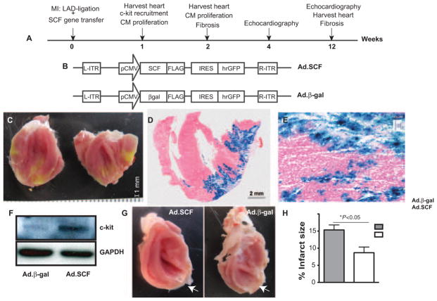
Objective: To determine whether stem cell factor gene transfer mediates cardiac regeneration in a rat myocardial infarction model, through survival and recruitment of c-kit(+) progenitors and cell-cycle activation in cardiomyocytes, and explore the mechanisms involved.

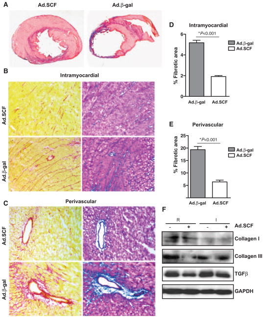
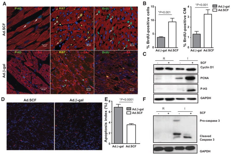
Methods and results: Infarct size, cardiac function, cardiac progenitor cells recruitment, fibrosis, and cardiomyocyte cell-cycle activation were measured at different time points in controls (n=10) and upon stem cell factor gene transfer (n=13) after myocardial infarction. We found a regenerative response because of stem cell factor overexpression characterized by an enhancement in cardiac hemodynamic function: an improvement in survival; a reduction in fibrosis, infarct size and apoptosis; an increase in cardiac c-kit(+) progenitor cells recruitment to the injured area; an increase in cardiomyocyte cell-cycle activation; and Wnt/β-catenin pathway induction.

Conclusions: Stem cell factor gene transfer induces c-kit(+) stem/progenitor cell expansion in situ and cardiomyocyte proliferation, which may represent a new therapeutic strategy to reverse adverse remodeling after myocardial infarction.


Figures

Figure 1
Figure 1. Study design
A, Experimental timeline. Our strategy consisted of using stem cell factor (SCF) as a therapy in cardiac regeneration in rats undergoing myocardial infarction (MI) after delivering adenoviruses expressing SCF membrane-bound form and green fluorescent protein (Ad.SCF) or adenoviruses containing β-gal and green fluorescent protein (Ad.β-gal) recombinant adenoviruses in the peri-infarct area. Myocardial regeneration and function was assessed at 1, 2, 4, and 12 weeks post-MI. B, Recombinant Ad.SCF and Ad.β-gal adenoviruses were generated after cloning SCF membrane isoform and β-gal gene sequences, respectively, into an adenoviral plasmid containing GFP as a reporter gene. CE, Detection of gene transfer within the myocardium 1 week post-MI. Distribution of the viral infection was confirmed by GFP expression, imaged through digital photography (C) and by X-gal staining (blue) (D and E) in control rats (Ad.β-gal). F, Analysis of c-kit receptor expression by western blot (WB), in lysates from control and Ad.SCF-treated hearts. α-GAPDH was used as protein loading control. G and H, Infarct size (white arrows) (G) was measured by cardiac magnetic resonance imaging 2 weeks post-MI (15.3±1.4% Ad.β-gal vs 8.6±1.6% Ad.SCF; *P<0.05; H). LAD indicates left anterior descending coronary artery; IRES, internal ribosomal entry site.
Figure 2
Figure 2. Cardiac function in the stem cell factor (SCF)-treated myocardium
A, Kaplan–Meier survival analysis in sham (n=5), adenoviruses containing β-gal and green fluorescent protein (Ad.β-gal)–treated group (n=10), and adenoviruses expressing SCF membrane-bound form and green fluorescent protein (Ad.SCF)-treated groups (n=13). Although there was no statistical significance, a tendency toward an increase is detected in survival after SCF treatment compared with controls 3 months postmyocardial infarction (MI; 90% Ad.SCF vs 65% Ad.β-gal; P=0.07 vs Ad.β-gal). BE, Echocardiographic measurements performed 1 and 3 months post-MI. Representative parameters short-axis M-mode view, in Ad.SCF-treated and control (Ad.β-gal) rats at 1 (B) and 3 months post-MI (C). Fractional shortening (FS) and ejection fraction (EF) 1 month (D) and 3 months (E) after Ad.SCF overexpression.
Figure 3
Figure 3. Cardiac fibrosis is decreased after stem cell factor (SCF) overexpression
A, Representative Masson trichrome–stained myocardial sections from control and SCF-treated rats 1 month post-myocardial infarction (MI). Blue, scar tissue; red, viable myocardium. BE, Intramyocardial (B) and periovascular fibrosis (C) analyzed by Picrosirius Red and Masson Trichrome staining 1 month post-MI. D and E, Effect of SCF overexpression in intramyocardial (D) (5.1%±0.2% in adenoviruses containing β-gal and green fluorescent protein [Ad.β-gal] vs 2.0%±0.1% in adenoviruses expressing SCF membrane-bound form and green fluorescent protein [Ad.SCF]; P<0.0001) and perivascular fibrosis (E) (19.3%±1.2% in Ad.β-gal vs 6.4%±0.7% in Ad.SCF; P<0.001). F, Analysis of collagen I, III, and transforming growth factor-β (TGF-β) expression by WB in cardiac lysates from remote (R) and infarcted (I) regions 3 months post-MI.
Figure 4
Figure 4. Cardiac c-kit+ population is increased at 1 week post-myocardial infarction (MI)
A, Confocal images represent c-kit membrane staining in section hearts. B, A 4-fold increase in c-kit+ cells is detected after stem cell factor (SCF) therapy. C, Cardiac c-kit+ cells detection by fluorescence-activated cell (FACS) sorting. D, Analysis of hematopoietic stem cell population (Lin c-kit+) by FACS in bone marrow and circulating blood cells shows no changes because of SCF overexpression. E and F, Cardiac c-kit+ population is gated for c-kit marker and early cardiac markers (GATA4, MEF2, MEF2C, and Nkx2.5) (E) or differentiated expression markers (eosinophils and high affinity IgE receptor [FcεRIα], multidrug resistant protein (MDR)-1, CD45, and α-sarcomeric actin [α-SA]) (F) are analyzed by FACS. G, Confocal images represent c-kit and CD45 membrane costaining in SCF-treated hearts. Ad.SCF indicates adenoviruses expressing SCF membrane-bound form and green fluorescent protein; Ad.β-gal, adenoviruses containing β-gal and green fluorescent protein.
Figure 5
Figure 5. Stem cell factor (SCF) therapy induces cardiomyocyte cell-cycle activation 1 week post-MI
A, Cardiomyocytes cell-cycle reentry (arrows) analyzed by costaining with α-sarcomeric actin (α-SA) and phosphohistone 3 (P-H3) or Ki67 proliferative markers. DNA synthesis is detected by bromodeoxyuridine (BrdU) incorporation. B, An increase of BrdU+ cells (10.1±1.4% adenoviruses expressing SCF membrane-bound form and green fluorescent protein [Ad.SCF] vs 3.7±0.3% adenoviruses containing β-gal and green fluorescent protein [Ad.β-gal]; *P<0.001; left) and BrdU+α-SA+ cells (CM) (3.8±0.3% Ad.SCF vs 1.8±0.2% Ad.β-gal, *P<0.001; right) is shown in the border area after SCF therapy. C, Cyclin D1, proliferating cell nuclear Ag (PCNA), and P-H3 proliferation expression markers in lysates from remote (R) and infarcted (I) regions. DF, SCF prevent apoptosis in the infarcted myocardium. Apoptotic cells detection by terminal deoxynucleotidyl transferase dUTP nick end labeling staining (red) in the border area (D). Apoptotic index (%) analysis by TUNEL (E) (7.0%±0.5% Ad.β-gal vs 3.5%±2.1% Ad.SCF; *P<0.0001). Caspase 3 activity analysis by western blot (WB) (F).
Figure 6
Figure 6. Proposed mechanism: Wnt/β-catenin pathway is activated in adenoviruses expressing stem cell factor (SCF) membrane-bound form and green fluorescent protein (Ad.SCF)-treated rats
A, β-catenin accumulation is observed in the remote (R) and infarcted (I) area 2 weeks after SCF overexpression. B, RNA isolated from remote (R) and infarcted (I) regions from controls and Ad.SCF-treated rats was reverse transcribed and Notch1, HoxB4, and cyclin D1 expression analyzed by quantitative real-time polymerase chain reaction. An upregulation of Notch1, HoxB4, and cyclin D1 is detected 2 weeks after SCF therapy. C, β-catenin and phospho-GSK3β increased expression is detected in isolated cardiomyocytes after SCF treatment. D, After myocardial infarction (MI), SCF therapy promotes cardiac repair by enhancing survival and left ventricle (LV) function and by preventing remodeling and apoptosis of existing cardiac cells. We hypothesize that SCF overexpression through c-kit receptor activation induces Wnt signaling activation in a paracrine way, promoting cardiac progenitors cells (CPCs) recruitment and cardiomyocytes (CM) cell-cycle activation and survival in the ischemic myocardium. RFU indicates relative fluorescence units.

Comment in

References

    1. Bergmann O, Bhardwaj RD, Bernard S, Zdunek S, Barnabé-Heider F, Walsh S, Zupicich J, Alkass K, Buchholz BA, Druid H, Jovinge S, Frisén J. Evidence for cardiomyocyte renewal in humans. Science. 2009;324:98–102. - PMC - PubMed
    1. Lutz M, Rosenberg M, Kiessling F, Eckstein V, Heger T, Krebs J, Ho AD, Katus HA, Frey N. Local injection of stem cell factor (SCF) improves myocardial homing of systemically delivered c-kit + bone marrow-derived stem cells. Cardiovasc Res. 2008;77:143–150. - PubMed
    1. Zsebo KM, Williams DA, Geissler EN, Broudy VC, Martin FH, Atkins HL, Hsu RY, Birkett NC, Okino KH, Murdock DC. Stem cell factor is encoded at the Sl locus of the mouse and is the ligand for the c-kit tyrosine kinase receptor. Cell. 1990;63:213–224. - PubMed
    1. Ayach BB, Yoshimitsu M, Dawood F, Sun M, Arab S, Chen M, Higuchi K, Siatskas C, Lee P, Lim H, Zhang J, Cukerman E, Stanford WL, Medin JA, Liu PP. Stem cell factor receptor induces progenitor and natural killer cell-mediated cardiac survival and repair after myocardial infarction. Proc Natl Acad Sci USA. 2006;103:2304–2309. - PMC - PubMed
    1. Smith MA, Pallister CJ, Smith JG. Stem cell factor: biology and relevance to clinical practice. Acta Haematol. 2001;105:143–150. - PubMed

Publication types

MeSH terms