. 2012 Aug 30;488(7413):642-6.
doi: 10.1038/nature11399.

Mutations in DMRT3 affect locomotion in horses and spinal circuit function in mice

Affiliations

Mutations in DMRT3 affect locomotion in horses and spinal circuit function in mice

Lisa S Andersson et al. Nature. .

Abstract

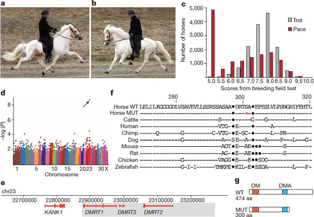
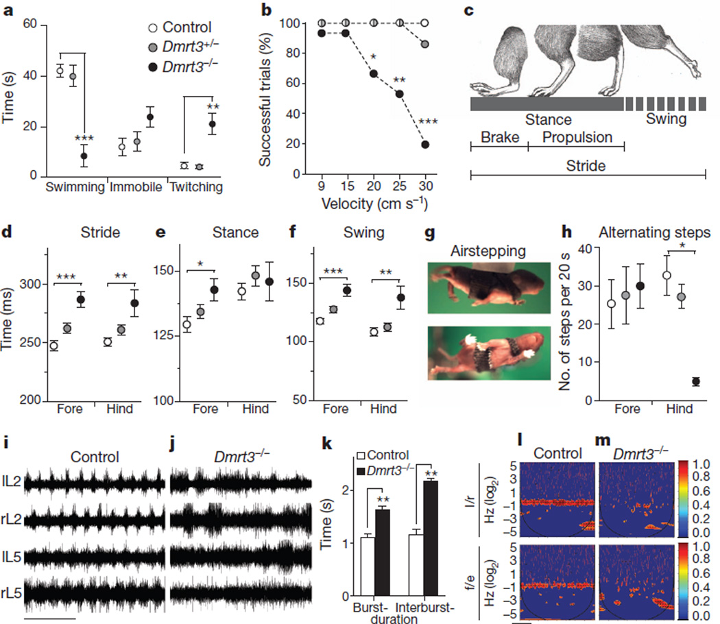
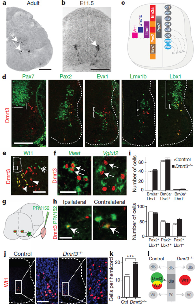
Locomotion in mammals relies on a central pattern-generating circuitry of spinal interneurons established during development that coordinates limb movement. These networks produce left-right alternation of limbs as well as coordinated activation of flexor and extensor muscles. Here we show that a premature stop codon in the DMRT3 gene has a major effect on the pattern of locomotion in horses. The mutation is permissive for the ability to perform alternate gaits and has a favourable effect on harness racing performance. Examination of wild-type and Dmrt3-null mice demonstrates that Dmrt3 is expressed in the dI6 subdivision of spinal cord neurons, takes part in neuronal specification within this subdivision, and is critical for the normal development of a coordinated locomotor network controlling limb movements. Our discovery positions Dmrt3 in a pivotal role for configuring the spinal circuits controlling stride in vertebrates. The DMRT3 mutation has had a major effect on the diversification of the domestic horse, as the altered gait characteristics of a number of breeds apparently require this mutation.


Figures

Figure 1
Figure 1. Identification of a DMRT3 mutation in horses
a, A pacing Icelandic horse, fore- and hindlegs on the same side of the body are synchronized. b, A trotting Icelandic horse, the diagonal fore- and hindlegs are synchronized. c, Distribution of breeding evaluation test scores for pace and trot in Icelandic horses. Score 5.0 indicates ‘gait not shown’. d, Genome-wide association analysis revealed a highly significant association between the ability to pace and SNP BIEC2_620109 on chromosome 23 (Praw = 1.7 × 10⁻⁹, corrected empirical P-value (EMP2) = 2.0 × 10⁻⁴, genome-wide significance). e, The 684 kb genomic interval associated with the Gait locus; the minimum Gait IBD region (438 kb) is shaded. f, Partial amino acid alignment of the predicted Dmrt3 protein in wild-type (WT) and mutant (MUT) horses and in other vertebrates. Horse nonsense mutation (red asterisk), sequence identities (dashes), insertions/deletions (dots). g, Schematics of wild-type and mutant Dmrt3. DM, zinc-finger like DNA binding module; DMA, protein domain of unknown function present in DMRT proteins.
Figure 2
Figure 2. Characterization of motor coordination in mice lacking Dmrt3
a, Dmrt3−/− mice showed decreased swimming duration (P < 0.0001), and an increase in twitching movements compared to control and Dmrt3+/− (P=0.0002); n = 5 control and Dmrt3−/−, n = 7 Dmrt3+/−. Time spent immobile was similar between genotypes (P=0.13). b, Mice were tested for their ability to run (>5 step cycles) at different treadmill speeds (9, 15, 20, 25, 30cm s−1) (n=15 trials per genotype, five animals). Dmrt3−/− mice had difficulties at 20cm s−1 (P=0.04) with markedly reduced number of successful trials at 25cm s−1 (P=0.006) and 30cm s−1 (P<0.0001) compared to controls. c, Schematic drawing of the gait parameters analysed in treadmill locomotion. df, At 20 cms−1 Dmrt3−/− showed increased stride (d, forelimb: P<0.0001, hindlimb: P = 0.006), hind limb stance (e, P = 0.02) and swing (f, forelimb: P<0.0001, hindlimb: P = 0.001) time duration compared to control (n = 11–12 trials per genotype, five animals). g, Images of a P4 mouse airstepping. h, Dmrt3−/− mice showed a decreased number of alternating hindlimb steps during airstepping (P=0.02 compared to controls; n=4 per genotype, except controls n=3). Wild-type littermate controls (white), Dmrt3+/− (grey), Dmrt3−/− (black). Mean ± s.e.m. im, Fictive locomotion was recorded from ventral left (l) and right (r) lumbar (L) root 2 and 5 from neonatal spinal cords. i, j, Representative traces from control (i) and Dmrt3−/− (j) spinal cords. Time scale 10 s. k, Dmrt3−/− mice displayed increased burst and interburst durations compared to control animals, analysis made on L2s (n = 6 per genotype, P = 0.02, burst; P = 0.001, interburst). l, m, Coherence power spectra analysis of left/right (l/r) and flexor/extensor (f/e) recordings (colour-graded scale; n=3–5 per genotype). Time scale 100 s.
Figure 3
Figure 3. Characterization of Dmrt3-expressing cells in the mouse spinal cord
a, Dmrt3 mRNA expression pattern in adult spinal cord (P60). b, Dmrt3 mRNA expression in a restricted population of neurons migrating ventrally in the developing spinal cord at E11.5. c, A schematic spinal cord cross-section showing progenitor and transcription factor domains. d, Double immunolabelling of Dmrt3 and Pax7 shows that Dmrt3+ cells originate from the ventral-most part (bracket) of the dorsal domain (border indicated by line). Dmrt3+ cells overlap with the dI4/dI6/V0d marker Pax2, but not with the V0V/V0C/V0G marker Evx1 or the dI5 marker Lmx1b (compare brackets). Dmrt3+ cells overlap with the dI4/dI5/dI6 marker Lbx1. e, Double immunolabelling with Dmrt3 (arrowhead) and Wt1 (double arrow) show a partial overlap (arrow). f, Dmrt3+ interneurons (arrows) co-labelled with Viaat mRNA (green) but not with Vglut2 mRNA (green). g, Schematic of trans-synaptic muscle tracing of Dmrt3 neurons in the spinal cord (n = 6, ipsilateral; n = 5, contralateral). h, Double immunolabelling of Dmrt3 and green fluorescent protein (PRV152) show that both ipsi- and contralateral premotor interneurons overlap with Dmrt3+ cells (arrows). i, Quantification of Brn3a/Lbx1-positive neurons (control nsection = 16, Dmrt3−/− nsection = 21) and of Lbx1/Pax2-positive neurons (control nsection = 11, Dmrt3−/− nsection=19). j, Immunolabelling of Wt1+ cells (red) in spinal cord sections from control and Dmrt3−/− E15.5 embryos. k, Quantification demonstrated that loss of Dmrt3 leads to an expanded Wt1+ cell subpopulation (control n=43, Dmrt3+/− n=36, Dmrt3−/− n=46, ***P<0.0001). l, Schematic illustration of fate change in the Dmrt3 dI6 population of neurons. Mean ± s.e.m. Scale bars: 400 µm (a), 70 µm (b, d, j), 50 µm (e, f, h).

References

    1. Grillner S. Biological pattern generation: the cellular and computational logic of networks in motion. Neuron. 2006;52:751–766. - PubMed
    1. Kullander K. Genetics moving to neuronal networks. Trends Neurosci. 2005;28:239–247. - PubMed
    1. Albertsdóttir E, Eriksson S, Sigurdsson A, Arnason T. Genetic analysis of ‘breeding field test status’ in Icelandic horses. J. Anim. Breed. Genet. 2011;128:124–132. - PubMed
    1. Hong CS, Park BY, Saint-Jeannet JP. The function of Dmrt genes in vertebrate development: it is not just about sex. Dev. Biol. 2007;310:1–9. - PubMed
    1. Ahituv N, et al. Deletion of ultraconserved elements yields viable mice. PLoS Biol. 2007;5:e234. - PMC - PubMed

Publication types

MeSH terms

Associated data