. 2012 Nov 1;303(9):E1107-16.
doi: 10.1152/ajpendo.00207.2012. Epub 2012 Aug 28.

SSTR2 is the functionally dominant somatostatin receptor in human pancreatic β- and α-cells

Affiliations

SSTR2 is the functionally dominant somatostatin receptor in human pancreatic β- and α-cells

Balrik Kailey et al. Am J Physiol Endocrinol Metab. .

Abstract

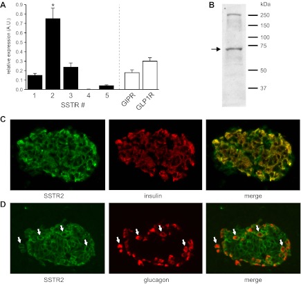
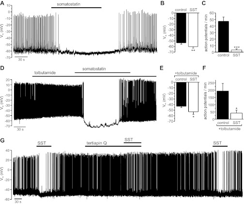
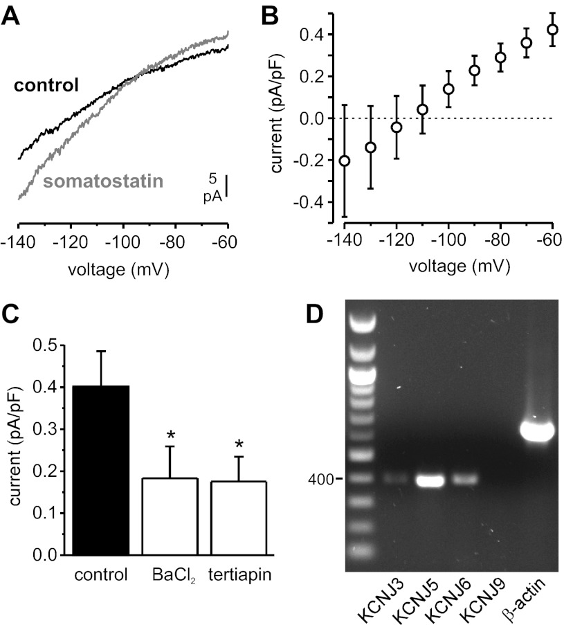
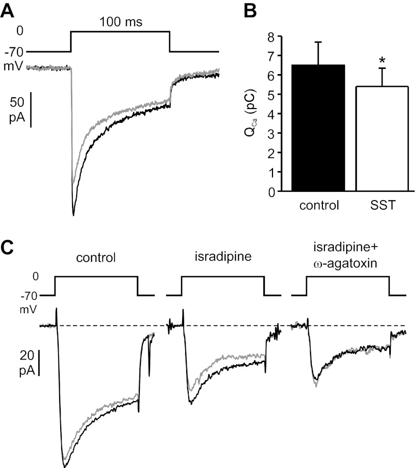
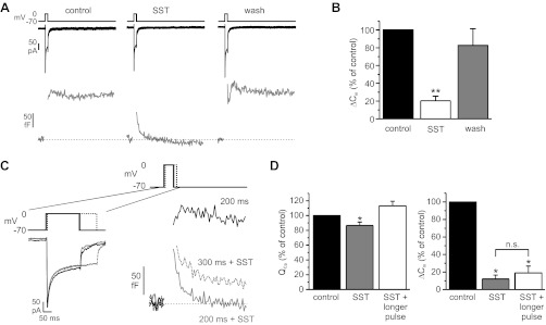
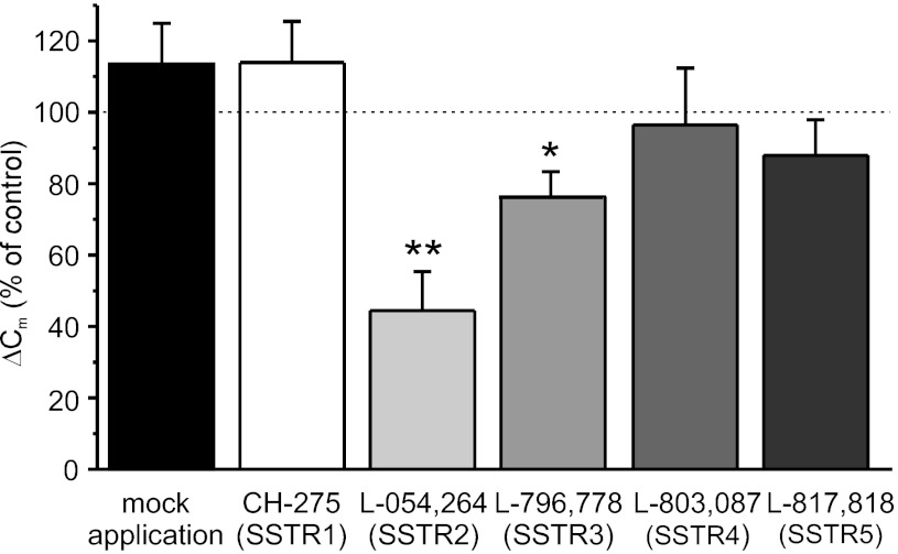
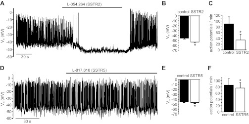
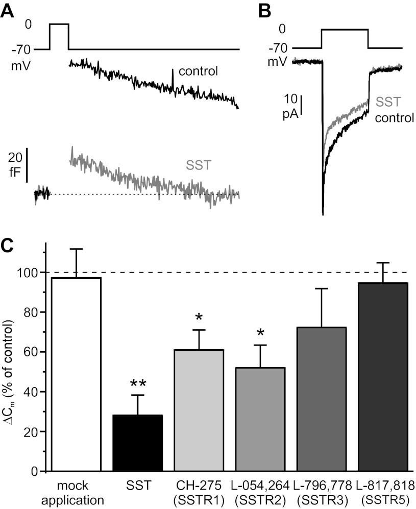
Somatostatin-14 (SST) inhibits insulin and glucagon secretion by activating G protein-coupled somatostatin receptors (SSTRs), of which five isoforms exist (SSTR1-5). In mice, the effects on pancreatic β-cells are mediated by SSTR5, whereas α-cells express SSTR2. In both cell types, SSTR activation results in membrane hyperpolarization and suppression of exocytosis. Here, we examined the mechanisms by which SST inhibits secretion from human β- and α-cells and the SSTR isoforms mediating these effects. Quantitative PCR revealed high expression of SSTR2, with lower levels of SSTR1, SSTR3, and SSTR5, in human islets. Immunohistochemistry showed expression of SSTR2 in both β- and α-cells. SST application hyperpolarized human β-cells and inhibited action potential firing. The membrane hyperpolarization was unaffected by tolbutamide but antagonized by tertiapin-Q, a blocker of G protein-gated inwardly rectifying K⁺ channels (GIRK). The effect of SST was mimicked by an SSTR2-selective agonist, whereas a SSTR5 agonist was marginally effective. SST strongly (>70%) reduced depolarization-evoked exocytosis in both β- and α-cells. A slightly weaker inhibition was observed in both cell types after SSTR2 activation. SSTR3- and SSTR1-selective agonists moderately reduced the exocytotic responses in β- and α-cells, respectively, whereas SSTR4- and SSTR5-specific agonists were ineffective. SST also reduced voltage-gated P/Q-type Ca²⁺ currents in β-cells, but normalization of Ca²⁺ influx to control levels by prolonged depolarizations only partially restored exocytosis. We conclude that SST inhibits secretion from both human β- and α-cells by activating GIRK and suppressing electrical activity, reducing P/Q-type Ca²⁺ currents, and directly inhibiting exocytosis. These effects are mediated predominantly by SSTR2 in both cell types.


Figures

Fig. 1.
Fig. 1.
Distribution of somatostatin receptor (SSTR) isoforms in human islets. A: analysis of relative SSTR isoform expression by quantitative RT-PCR in 10 human islet preparations. Data are displayed relative to standard housekeeping genes [peptidylprolyl isomerase A (PPIA) and ubiquitin C (UBC)] using the ΔCT method. SSTR2 relative expression is significantly (P < 0.05) higher than the other SSTR isoforms as well as 2 other islet-expressed G protein-coupled receptors (GPCRs) [gastric inhibitory polypeptide receptor (GIPR) and glucagon-like peptide-1 receptor (GLP-1R)]. B: Western blot analysis of isolated human islets using a SSTR2-specific antibody (expected molecular weight: ∼72 kDa). Positions of molecular weight markers are shown on the right. C and D: coimmunostaining of human pancreatic tissue sections with anti-SSTR2 and anti-insulin (C) or anti-glucagon (D). White arrows indicate cells positive for both SSTR2 and glucagon. AU, arbitrary units.
Fig. 2.
Fig. 2.
Effect of somatostatin (SST) on the β-cell membrane potential (Vm). All experiments were performed in the presence of 6 mM glucose. A: SST (20 nM) was applied to an electrically active β-cell as indicated. B and C: average effect of SST (10–30 nM) on Vm (n = 7; **P < 0.01) and action potential frequency (n = 6; ***P < 0.001). D: SST (30 nM) was applied in the presence of tolbutamide (200 μM), as indicated by the bars. E and F: average effect of SST (30 nM) on Vm (n = 6) and action potential frequency (n = 4) when applied in the presence of tolbutamide (200 μM). *P < 0.05. G: SST (30 nM) was applied in the absence or presence of tertiapin-Q (100 nM), as indicated by bars.
Fig. 3.
Fig. 3.
Effect of SST on the β-cell resting membrane current (in the presence of 6 mM extracellular glucose). A: representative recording of membrane currents elicited by voltage ramps from −140 to −60 mV (speed: 0.2 V/s) before (black trace) and during application of 30 nM SST (gray trace). B: current-voltage relationship of the SST-activated current (obtained by subtracting traces recorded before SST application from traces recorded during SST application and normalized to cell size; n = 5). C: amplitude of the outward current at −70 mV (normalized to cell size) activated by SST application in the absence (control) or presence of Ba2+ (1 mM) or tertiapin-Q (0.3 μM; n = 5 in each group). *P < 0.05 vs control. D: RT-PCR analysis of expression of G protein-gated inwardly rectifying K+ channel subunits in human islets (the 400-bp marker is indicated on the left). KCNJ3, Kir3.1; KCNJ5, Kir3.4; KCNJ6, Kir3.2; KCNJ9, Kir3.3.
Fig. 4.
Fig. 4.
Effect of SST on β-cell voltage-gated Ca2+ currents. A: representative recording showing Ca2+ currents evoked by 100-ms depolarizations from −70 to 0 mV before (black trace) and during application of 30 nM SST (gray trace) in the same cell. B: the average effect on the integrated Ca2+ current (QCa) from 6 experiments is shown (*P < 0.05). C: SST (30 nM) was applied to the same cell under control conditions (left), in the presence of 10 μM isradipine (middle), and in the combined presence of isradipine and 200 nM ω-agatoxin IVA (right). Black traces are before and gray traces during SST application (50-ms depolarizations). The effect of SST was reversible after each application (not shown).
Fig. 5.
Fig. 5.
Effect of SST on β-cell exocytosis and involvement of Ca2+ current. A: inward Ca2+ current (black trace) and capacitance response (gray trace) evoked by 300-ms depolarizations from −70 to 0 mV before (left) and during exposure to 30 nM SST (middle) and after removal of the hormone (right). B: summary of capacitance responses (ΔCm) from 13 cells (**P < 0.01). C: Ca2+ currents (left) and capacitance responses (right) evoked by a 200-ms depolarization under control conditions (black traces), by a 200-ms depolarization in the presence of 30 nM SST (gray traces), and by a 300-ms depolarization in the continued presence of SST (dotted lines) in the same cell. D: integrated Ca2+ currents (QCa) and exocytotic responses (ΔCm) from 4 experiments performed similarly as described in C (i.e., the Ca2+ influx in the presence of SST was increased above pre-SST levels by prolongation of the depolarizing pulse). *P < 0.05.
Fig. 6.
Fig. 6.
Effects of subtype-selective SSTR agonists on depolarization-evoked exocytosis (ΔCm) in β-cells (n = 7, 6, 10, 8, 5, and 7 in mock application, CH-275, L-054,264, L-796–778, L-803–087, and L-817–818, respectively). *P < 0.05; **P < 0.01.
Fig. 7.
Fig. 7.
Effects of SSTR2- and SSTR5-selective agonists on β-cell electrical activity. A and D: representative recordings; agonists were applied as indicated by bars. Average effects on Vm (B and E) and action potential frequency (C and F); n = 8 (SSTR2) and 5 (SSTR5). *P < 0.05.
Fig. 8.
Fig. 8.
Effects of SST and SSTR subtype-specific agonists on α-cell exocytosis and Ca2+ current. Representative recordings of capacitance responses (A) and Ca2+ currents (B) evoked by 500-ms depolarizing pulses from −70 to 0 mV under control conditions (black traces) and in the presence of 30 nM SST (gray traces). C: average effects of SST and SSTR agonists on exocytotic responses (ΔCm; n = 6, 8, 8, 5, 8, and 5 in mock application, SST, CH-275, L-054–264, L-796–778, and L-817,818, respectively). *P < 0.05; **P < 0.01.

References

    1. Barnett P. Somatostatin and somatostatin receptor physiology. Endocrine 20: 255–264, 2003 - PubMed
    1. Ben-Shlomo A, Melmed S. Pituitary somatostatin receptor signaling. Trends Endocrinol Metab 21: 123–133, 2010 - PMC - PubMed
    1. Bosco D, Armanet M, Morel P, Niclauss N, Sgroi A, Muller YD, Giovannoni L, Parnaud G, Berney T. Unique arrangement of alpha- and beta-cells in human islets of Langerhans. Diabetes 59: 1202–1210, 2010 - PMC - PubMed
    1. Braun M, Ramracheya R, Amisten S, Bengtsson M, Moritoh Y, Zhang Q, Johnson PR, Rorsman P. Somatostatin release, electrical activity, membrane currents and exocytosis in human pancreatic delta cells. Diabetologia 52: 1566–1578, 2009 - PubMed
    1. Braun M, Ramracheya R, Bengtsson M, Clark A, Walker JN, Johnson PR, Rorsman P. Gamma-aminobutyric acid (GABA) is an autocrine excitatory transmitter in human pancreatic beta-cells. Diabetes 59: 1694–1701, 2010 - PMC - PubMed

Publication types

MeSH terms

LinkOut - more resources