. 2013 Sep;218(5):1115-32.
doi: 10.1007/s00429-012-0450-1. Epub 2012 Aug 30.

Kv4.2 potassium channels segregate to extrasynaptic domains and influence intrasynaptic NMDA receptor NR2B subunit expression

Affiliations

Kv4.2 potassium channels segregate to extrasynaptic domains and influence intrasynaptic NMDA receptor NR2B subunit expression

Walter A Kaufmann et al. Brain Struct Funct. 2013 Sep.

Abstract

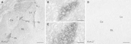
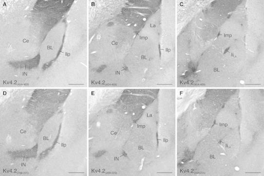
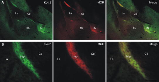
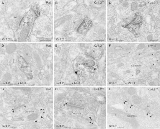
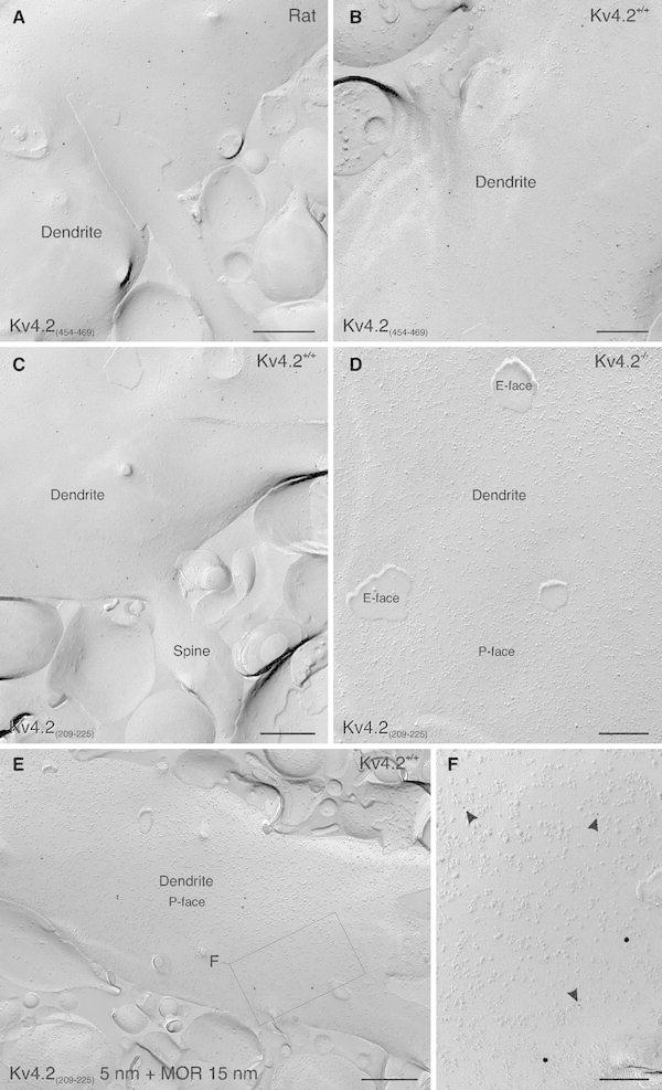
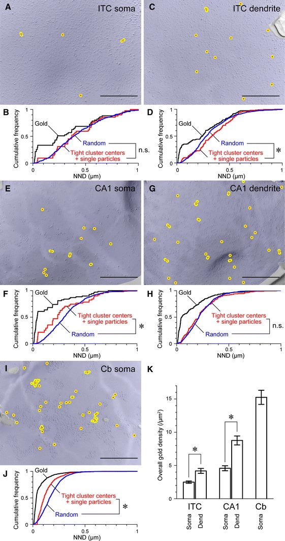
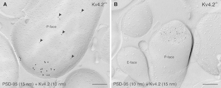
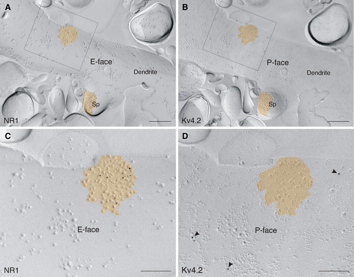
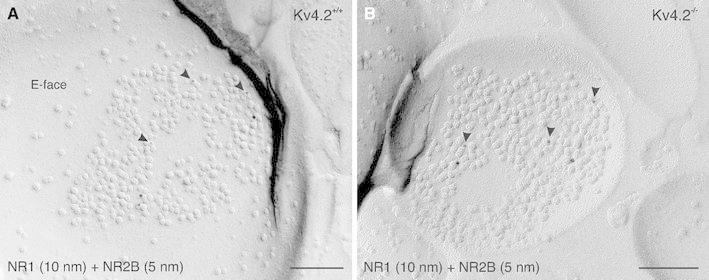
Neurons of the intercalated cell clusters (ITCs) represent an important relay site for information flow within amygdala nuclei. These neurons receive mainly glutamatergic inputs from the basolateral amygdala at their dendritic domains and provide feed-forward inhibition to the central nucleus. Voltage-gated potassium channels type-4.2 (Kv4.2) are main players in dendritic signal processing and integration providing a key component of the A currents. In this study, the subcellular localization and distribution of the Kv4.2 was studied in ITC neurons by means of light- and electron microscopy, and compared to other types of central principal neurons. Several ultrastructural immunolocalization techniques were applied including pre-embedding techniques and, most importantly, SDS-digested freeze-fracture replica labeling. We found Kv4.2 densely expressed in somato-dendritic domains of ITC neurons where they show a differential distribution pattern as revealed by nearest neighbor analysis. Comparing ITC neurons with hippocampal pyramidal and cerebellar granule cells, a cell type- and domain-dependent organization in Kv4.2 distribution was observed. Kv4.2 subunits were localized to extrasynaptic sites where they were found to influence intrasynaptic NMDA receptor subunit expression. In samples of Kv4.2 knockout mice, the frequency of NR1-positive synapses containing the NR2B subunit was significantly increased. This indicates a strong, yet indirect effect of Kv4.2 on the synaptic content of NMDA receptor subtypes, and a likely role in synaptic plasticity at ITC neurons.


Figures

Fig. 1
Fig. 1
Distribution of Kv4.2 in the mouse amygdala revealed by immunoperoxidase labeling. a Prominent Kv4.2 immunoreactivity is detected in all ITC clusters and the ITC nucleus in C57Bl/6 wild-type mice (Kv4.2+/+). Only low to moderate levels of immunoreactivity are observed in other amygdaloid areas than the ITC. b Higher magnification of the Imp cluster (boxed area in a). Diffuse Kv4.2 immunoreactivity is primarily observed in the neuropil. c A similar immunostaining pattern for Kv4.2 is observed in the ITC nucleus (boxed area in a). d Specificity of Kv4.2 immunolabeling is confirmed on respective brain areas from a Kv4.2−/− mouse. BL basolateral amygala, Ce central nucleus of amygdala, Ilp lateral paracapsular ITC cluster, Imp medial paracapsular ITC cluster, IN ITC nucleus, Is LA supralateral ITC cluster, La lateral amygdala. Scale bar 200 μm (a, d); 40 μm (b, c)
Fig. 2
Fig. 2
Kv4.2 immunolabeling in the rat amygdala using antibodies directed against different regions of the Kv4.2 protein. ac The Kv4.2(454–469) antibody reveals prominent immunolabeling of all ITC clusters and the ITC nucleus from rostral (a) to medial (b) and caudal (c) levels of the amygdala using an immunoperoxidase staining technique. Other amygdaloid areas are only faintly labeled. df An equivalent immunolabeling pattern is observed with the Kv4.2(209–225) antibody. BL basolateral amygdala, Ce central nucleus of amygdala, Ii LA intralateral paracapsular ITC cluster, Ilp lateral paracapsular ITC cluster, Imp medial paracapsular ITC cluster, IN ITC nucleus, La lateral nucleus of amygdala. Scale bar 350 μm (af)
Fig. 3
Fig. 3
Colocalization of Kv4.2 and μ-opioid receptors (MOR) in the ITCs. a Double-labeling immunofluorescence for Kv4.2 (in green) and MOR (in red) reveals a high degree of coexistence in the amygdala, which is evident in the merged images. b At higher resolution, immunoreactivity for Kv4.2 (in green) as well as for MOR (in red) appears dense and diffuse in ITC clusters such as the Imp. The distribution profile of these proteins is highly similar (merge). BL basolateral amygdala, Ce central nucleus of amygdala, Imp medial paracapsular ITC cluster, IN ITC nucleus, La lateral amygdala. Scale bar 350 μm (a), 50 μm (b)
Fig. 4
Fig. 4
Subcellular localization of Kv4.2 to somato-dendritic domains of ITC neurons revealed by pre-embedding immunoperoxidase and immunometal electron microscopy. a A small dendritic trunk of a neuron in the rat ITC nucleus displays dense and diffuse immunolabeling for Kv4.2 [Kv4.2(454–469) antibody]. The immunolabeling appears as an electron-opaque reaction product. A synaptic terminal, contacting this dendrite, is free of any immunolabeling. b A dendritic spine of a neuron in the mouse ITC nucleus shows dense and diffuse labeling for Kv4.2 [Kv4.2(209–225) antibody]. An axon terminal contacting this spine, as well as other cellular profiles, is free of any immunolabeling. c A dendritic spine of a mouse Imp neuron shows the same dense and diffuse labeling pattern for Kv4.2 [Kv4.2(454–469) antibody]. d In a double-labeling experiment, a Kv4.2 immunopositive spine of a rat Imp neuron (immunoperoxidase reaction) is also immunoreactive for MOR (immunometal reaction). e A small dendrite of a mouse Imp neuron, immunolabeled for Kv4.2 (immunoperoxidase reaction), is also immunoreactive for MOR (immunometal reaction). An axon terminal, contacting this dendrite, is free of any immunolabeling. f In a sample from a Kv4.2−/− mouse, a large dendrite of an Imp neuron is labeled for MOR (immunometal reaction), but devoid of Kv4.2 immunolabeling (immunoperoxidase reaction). g Immunometal particles (indicated by arrowheads) for Kv4.2, when applying the Kv4.2(454–469) antibody, are observed at the intracellular side of the plasma membrane of a rat Imp neuron. The postsynaptic specialization of an excitatory synapse is free of any immunolabeling. Some immunoparticles appear localized perisynaptically. h Immunometal particles (indicated by arrowheads) for Kv4.2 appear at the intracellular side of an ITC neuron dendrite also when applying the Kv4.2(209–225) antibody. Both the axon forming a synapse with this dendrite and the postsynaptic specialization are free of any immunolabeling. i The plasma membrane of an Imp dendrite is decorated with immunoparticles at the intracellular side applying the Kv4.2(209–225) antibody. The postsynaptic specialization of an inhibitory synapse on this dendrite is also free of any immunolabeling. At axon terminal, MOR μ-opioid receptor. Scale bar 200 nm (ac, e, h, i); 300 nm (d, f, g)
Fig. 5
Fig. 5
SDS-FRL confirms the localization of Kv4.2 in the somato-dendritic plasma membrane of ITC neurons. a Immunoparticles labeling Kv4.2 subunits (10 nm gold) are scattered on the plasma membrane of a large dendritic trunk of a rat ITC neuron. The immunolabeling is restricted to the plasma membrane P-face. b Scattered immunopartices are also observed in the plasma membrane P-face of a large dendritic trunk of a mouse ITC neuron. c In addition to the labeling of the dendritic trunk, Kv4.2 immunoparticles are localized to the head and neck of spines. d Both the plasma membrane E- and P-face of an ITC dendrite from a Kv4.2−/− mouse are immunonegative when reacted with the Kv4.2(209–225) antibody (10 nm gold). e An ITC dendritic trunk from a wild-type mouse (Kv4.2+/+) is immunopositive for both Kv4.2(209–225) (5 nm gold) and MOR (15 nm gold) in a double-immunolabeling experiment. f At higher resolution (boxed area in e), both 5 and 15 nm gold particles revealing Kv4.2 subunits and MOR, respectively, are visible on the P-face of the dendritic plasma membrane. MOR μ-opioid receptor. Scale bar 300 nm (a, c); 200 nm (b, d); 400 nm (e); 150 nm (f)
Fig. 6
Fig. 6
Quantitative characterization of the distribution of Kv4.2 immunogold particles in ITC neurons and other central principal cells. a A sample SDS-FRL image of a portion of an ITC neuron soma. The area of the relevant profile is colored in blue. Immunogold particles labeling Kv4.2 subunits are marked with a black dot, a 20-nm radius circle around each particle is shown in yellow and the centroid of overlapping circles is marked with a red open circle. A tight cluster of particles is defined as particles residing within the overlapping yellow circles. b Cumulative probability curves for the nearest neighbor distances (NNDs) between individual immunogold particles (in black) and tight cluster centers along with single particles (in red), and NNDs of the calculated random distribution (in blue). c A sample image of a portion of an ITC dendrite and d respective NND analysis. eh Sample images and NND analyses for CA1 pyramidal neuron soma and dendrite. ij Sample image and NND analysis for cerebellar granule cell soma. k Histogram showing the immunogold particle density in subcellular domains of ITC neurons (soma: 8 profiles with a mean area of 2.60 μm2; dendrite: 22 profiles with a mean area of 2.05 μm2), CA1 pyramidal neurons (soma: 6 profiles with a mean area of 2.52 μm2; dendrite: 19 profiles with a mean area of 1.52 μm2) and cerebellar granule cells (soma: 25 profiles with a mean area of 2.54 μm2). Cb cerebellar granule cell, NND nearest neighbor distance. Error bars, SEM. *p < 0.05, Mann–Whitney U. Scale bar 500 nm (a, c, e, g, i)
Fig. 7
Fig. 7
Segregation of Kv4.2 to extrasynaptic domains. a In a double-immunolabeling experiment for PSD-95, visualized with 15 nm gold particles, and Kv4.2, visualized with 10 nm gold particles (arrowheads), Kv4.2 subunits are observed at extrasynaptic sites of an ITC dendritic spine in a wild-type mouse (Kv4.2+/+). Conversely, PSD-95 labeling can be seen within the postsynaptic membrane specialization of an excitatory synapse. b In a Kv4.2−/− mouse, labeling for PSD-95 (10 nm gold) can clearly be detected within the postsynaptic membrane specialization of a dendritic spine. No immunoreactivity for Kv4.2 (15 nm gold) is observed in Kv4.2−/− mouse tissue. Scale bar 200 nm (a, b)
Fig. 8
Fig. 8
Kv4.2 does not reside within the postsynaptic membrane specializations of excitatory synapses as revealed by a double-replica approach. a Immunogold particles revealing NR1 subunits (10 nm gold) are localized to the E-face of an ITC dendrite. b On the mirror replica, immunogold particles revealing Kv4.2 subunits (10 nm gold) are observed on the P-face of the same dendrite. c At higher resolution (boxed area in a), immunolabeling for NR1 subunits is evident on the E-face of a postsynaptic membrane specialization of a glutamatergic synapse (shown in orange), which is characterized by the clustering of intramembrane particles. d Immunolabeling for Kv4.2 subunits is present on the P-face of the extrasynaptic plasma membrane at higher resolution (boxed area in b). The postsynaptic membrane specialization by itself is free of any Kv4.2 immunolabeling. Sp spine. Scale bar 300 nm (a, b); 150 nm (c, d)
Fig. 9
Fig. 9
Modulatory action of Kv4.2 on the synaptic expression of NR2B. a In a double-immunolabeling experiment for NMDA-type glutamate receptor subunit 1 (NR1) and subunit 2B (NR2B), both NR1 (10 nm gold) and NR2B (5 nm gold; arrowheads) are observed on the P-face of the postsynaptic membrane specialization of an excitatory synapse in an ITC neuron from a Kv4.2+/+ mouse. b In an ITC neuron from a Kv4.2−/− mouse, NR1 (10 nm gold) and NR2B immunolabeling (5 nm gold; arrowheads) are observed in the postsynaptic membrane of an excitatory synapse at a density similar to that observed in Kv4.2+/+ neurons. Yet, the frequency of such synapses, co-immunolabeled for both the NR1 and NR2B subunits, was increased in ITC neurons of Kv4.2−/− mice. Scale bar 150 nm (a, b)

Similar articles

Cited by

References

    1. Alonso G, Widmer H. Clustering of KV4.2 potassium channels in postsynaptic membrane of rat supraoptic neurons: an ultrastructural study. Neuroscience. 1997;77(3):617–621. doi: 10.1016/S0306-4522(96)00561-1. - DOI - PubMed
    1. An WF, Bowlby MR, Betty M, Cao J, Ling HP, Mendoza G, Hinson JW, Mattsson KI, Strassle BW, Trimmer JS, Rhodes KJ. Modulation of A-type potassium channels by a family of calcium sensors. Nature. 2000;403(6769):553–556. doi: 10.1038/35000592. - DOI - PubMed
    1. Arvidsson U, Riedl M, Chakrabarti S, Lee JH, Nakano AH, Dado RJ, Loh HH, Law PY, Wessendorf MW, Elde R. Distribution and targeting of a mu-opioid receptor (MOR1) in brain and spinal cord. J Neurosci. 1995;15(5 Pt 1):3328–3341. - PMC - PubMed
    1. Barria A, Malinow R. NMDA receptor subunit composition controls synaptic plasticity by regulating binding to CaMKII. Neuron. 2005;48(2):289–301. doi: 10.1016/j.neuron.2005.08.034. - DOI - PubMed
    1. Birnbaum SG, Varga AW, Yuan LL, Anderson AE, Sweatt JD, Schrader LA. Structure and function of Kv4-family transient potassium channels. Physiol Rev. 2004;84(3):803–833. doi: 10.1152/physrev.00039.2003. - DOI - PubMed

Publication types

MeSH terms

Substances

LinkOut - more resources