. 2012 Sep 18;109(38):15101-8.
doi: 10.1073/pnas.1213353109. Epub 2012 Aug 29.

Causes, consequences, and remedies for growth-induced solid stress in murine and human tumors

Affiliations

Causes, consequences, and remedies for growth-induced solid stress in murine and human tumors

Triantafyllos Stylianopoulos et al. Proc Natl Acad Sci U S A. .

Abstract

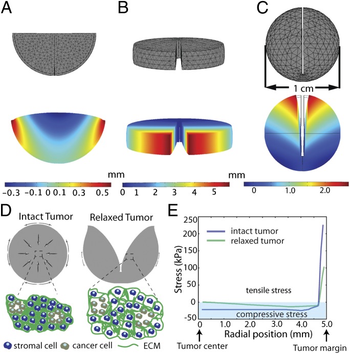
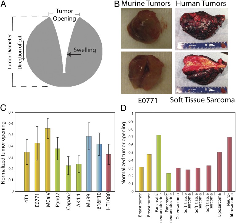
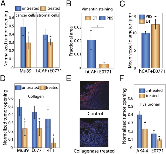
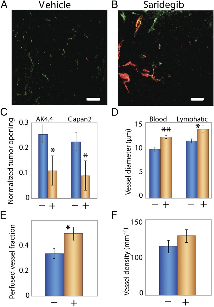
The presence of growth-induced solid stresses in tumors has been suspected for some time, but these stresses were largely estimated using mathematical models. Solid stresses can deform the surrounding tissues and compress intratumoral lymphatic and blood vessels. Compression of lymphatic vessels elevates interstitial fluid pressure, whereas compression of blood vessels reduces blood flow. Reduced blood flow, in turn, leads to hypoxia, which promotes tumor progression, immunosuppression, inflammation, invasion, and metastasis and lowers the efficacy of chemo-, radio-, and immunotherapies. Thus, strategies designed to alleviate solid stress have the potential to improve cancer treatment. However, a lack of methods for measuring solid stress has hindered the development of solid stress-alleviating drugs. Here, we present a simple technique to estimate the growth-induced solid stress accumulated within animal and human tumors, and we show that this stress can be reduced by depleting cancer cells, fibroblasts, collagen, and/or hyaluronan, resulting in improved tumor perfusion. Furthermore, we show that therapeutic depletion of carcinoma-associated fibroblasts with an inhibitor of the sonic hedgehog pathway reduces solid stress, decompresses blood and lymphatic vessels, and increases perfusion. In addition to providing insights into the mechanopathology of tumors, our approach can serve as a rapid screen for stress-reducing and perfusion-enhancing drugs.


Conflict of interest statement

Conflict of interest statement: R.K.J. received research grants from Dyax, MedImmune, and Roche; received consultant fees from Dyax, Enlight, Noxxon, and SynDevRx; owns equity in Enlight, SynDevRx, and XTuit; and serves on the Board of Directors of XTuit and Board of Trustees of H&Q Healthcare Investors and H&Q Life Sciences Investors. No reagents or funding from these companies were used in these studies. Therefore, there is no significant financial or other competing interest in the work.

Figures

Fig. 1.
Fig. 1.
Model predictions for the total displacement of the tumor after releasing the growth-induced stress by (A) cutting the tumor in one-half, (B) cutting a slice of the tumor, and (C) cutting the whole tumor. The tumor has a diameter of 1 cm, and the depth of the cut (B and C) is 0.8 cm. Notice the different displacement scales on the legends. Negative total displacements denote swelling (compressive stress), and positive total displacements denote opening (tensile stress) of the tissue. (D) Schematic of growth-induced stresses in tumors. In the tumor center, circumferential and radial stresses are compressive; in the periphery, radial stress is compressive, and circumferential stress is tensile (direction indicated with arrows). A partial cut through the center of the tumor (80% through the diameter) releases the stresses, and the tumor deforms in a measurable way. Compressive stresses in the tumor interior squeeze tumor components. After the tumor is cut and the stresses are released, the tumor interior decompresses (swells). (E) Spatial distribution of the circumferential growth-induced stress in an intact tumor and a tumor after making a cut. Compressive circumferential stresses at the tumor interior diminish, whereas tensile circumferential stresses at the periphery are also alleviated considerably.
Fig. 2.
Fig. 2.
Estimation of stress in transplanted and human tumors. (A) Schematic of tumor deformation after making a cut and releasing growth-induced stress. The retraction of the tumor at the surface is indicative of circumferential tension at the tumor margin, whereas the swelling of the inner surface at the point of cut is indicative of compression in the intratumoral region, which presumably balances the tension at the margin. Measuring the displacement (tumor opening) accounts for both retraction and swelling. (B) Photographs of the initial and final shape of tumors after making a cut. (C and D) Tumor openings were measured in transplanted orthotopic tumors surgically excised from mice and human tumors surgically excised from patients. The diameters of the mouse tumors (C) were ∼1 cm, and the dimensions of the human tumors (D) are given in Table S3. Normalized tumor opening is the tumor opening as a fraction of the initial tumor diameter. For nonspherical tumors, an equivalent diameter was used for a sphere that has the same volume as the volume of the tumors. Yellow columns represent breast tumors, green columns represent pancreatic tumors, blue columns represent melanomas, and maroon columns represent sarcomas.
Fig. 3.
Fig. 3.
Selective depletion of tumor constituents reduces growth-induced stress and decreases tumor opening. (A) Depletion with diphtheria toxin of human cancer cells in human Mu89 tumors or human stromal cells in E0771 tumors coimplanted with human CAFs decreased tumor opening (P = 0.001 and P = 0.023, respectively; Student t test). (B) Staining of E0771 tumors for colocalization of vimentin confirms that human fibroblast levels were reduced by diphtheria toxin treatment. The decrease in vimentin-stained area is significant (P = 0.03; Student t test). (C) Depletion of stromal cells also increased mean blood vessel diameter (P = 0.05; Student t test). (D) Treatment with bacterial collagenase decreased the tumor opening significantly and thus, the growth-induced stress for all tumors (P = 0.003 for Mu89, P = 0.025 for E0771, P = 4 × 10⁻⁶ for 4T1; Student t test). (E) Staining of Mu89 tumors for cell nuclei (blue) and collagen (red) shows reduction of collagen after treatment with collagenase. (F) Treatment with hyaluronidase also significantly decreased the tumor opening in AK4.4 and E0771 tumors (P = 0.022 and P = 0.027, respectively; Student t test). All tumor models are orthotopic. The asterisks denote a statistically significant difference.
Fig. 4.
Fig. 4.
Saridegib increases tumor vessel diameter and reopens compressed vessels by reducing stress. (A) Orthotopically transplanted AK4.4 pancreatic tumors are hypovascular with compressed vessel structures stained with CD31 (green). These tumors also have reduced perfusion, which was detected by lectin (red). (B) Treatment with saridegib increased the number of lectin-perfused vessels. (C–F) Treatment with saridegib reduced stress (C; P = 0.046 for AK4.4 and P = 0.022 for Capan2; Student t test) in tumors, leading to increased vessel diameter (D; blood vessels: CD31 staining, P = 0.003; lymphatic vessels: LYVE-1 staining, P = 0.004; Student t test) and fraction of perfused vessels (E; P = 0.049; Student t test) without increasing vessel density (F; P = not significant) in AK4.4. +, saridegib; −, vehicle. The asterisks denote a statistically significant difference. (Scale bar, 100 μm.)
Fig. 5.
Fig. 5.
Strategies to alleviate growth-induced solid stress in tumors. (Middle) In an untreated tumor, proliferating cancer cells and activated fibroblasts deform the ECM, resulting in stretched collagen fibers, compressed hyaluronan, and deformed cells—all storing solid stress. This stress compresses intratumor blood and lymphatic vessels. Potential strategies to alleviate solid stress and decompress vessels involve depleting these components. Depleting cancer cells (Top Left) or fibroblasts (Top Right) relaxes collagen fibers, hyaluronan, and the remaining cells, alleviating solid stress. Depleting collagen (Bottom Left) alleviates the stress that was held within these fibers as well as relaxes stretched/activated fibroblasts and compressed cancer cells within nodules. Finally, depleting hyaluronan (Bottom Right) alleviates the stored compressive stress, allowing nearby components to decompress. (Note that other stromal cells, such as pericytes, macrophages, and various immune cells that might also control production of collagen or hyaluronan, are not shown to simplify the schematic. Lymphatic vessels are also not shown for the same reason.)

References

    1. Jain RK. Normalization of tumor vasculature: An emerging concept in antiangiogenic therapy. Science. 2005;307:58–62. - PubMed
    1. Young JS, Lumsden CE, Stalker AL. The significance of the tissue pressure of normal testicular and of neoplastic (Brown-Pearce carcinoma) tissue in the rabbit. J Pathol Bacteriol. 1950;62:313–333. - PubMed
    1. Jain RK, Baxter LT. Mechanisms of heterogeneous distribution of monoclonal antibodies and other macromolecules in tumors: Significance of elevated interstitial pressure. Cancer Res. 1988;48:7022–7032. - PubMed
    1. Baxter LT, Jain RK. Transport of fluid and macromolecules in tumors. I. Role of interstitial pressure and convection. Microvasc Res. 1989;37:77–104. - PubMed
    1. Boucher Y, Baxter LT, Jain RK. Interstitial pressure gradients in tissue-isolated and subcutaneous tumors: Implications for therapy. Cancer Res. 1990;50:4478–4484. - PubMed

Publication types