

Prevalence of abnormalities in knees detected by MRI in adults without knee osteoarthritis: population based observational study (Framingham Osteoarthritis Study)
- PMID: 22932918
- PMCID: PMC3430365
- DOI: 10.1136/bmj.e5339
Prevalence of abnormalities in knees detected by MRI in adults without knee osteoarthritis: population based observational study (Framingham Osteoarthritis Study)
Abstract
Objective: To examine use of magnetic resonance imaging (MRI) of knees with no radiographic evidence of osteoarthritis to determine the prevalence of structural lesions associated with osteoarthritis and their relation to age, sex, and obesity.
Design: Population based observational study.
Setting: Community cohort in Framingham, MA, United States (Framingham osteoarthritis study).
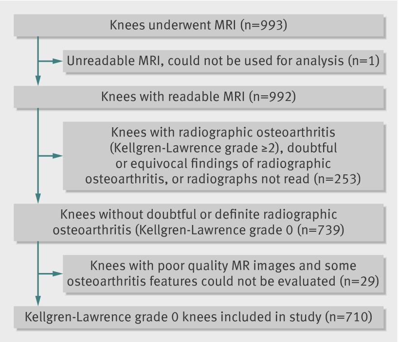
Participants: 710 people aged >50 who had no radiographic evidence of knee osteoarthritis (Kellgren-Lawrence grade 0) and who underwent MRI of the knee.
Main outcome measures: Prevalence of MRI findings that are suggestive of knee osteoarthritis (osteophytes, cartilage damage, bone marrow lesions, subchondral cysts, meniscal lesions, synovitis, attrition, and ligamentous lesions) in all participants and after stratification by age, sex, body mass index (BMI), and the presence or absence of knee pain. Pain was assessed by three different questions and also by WOMAC questionnaire.
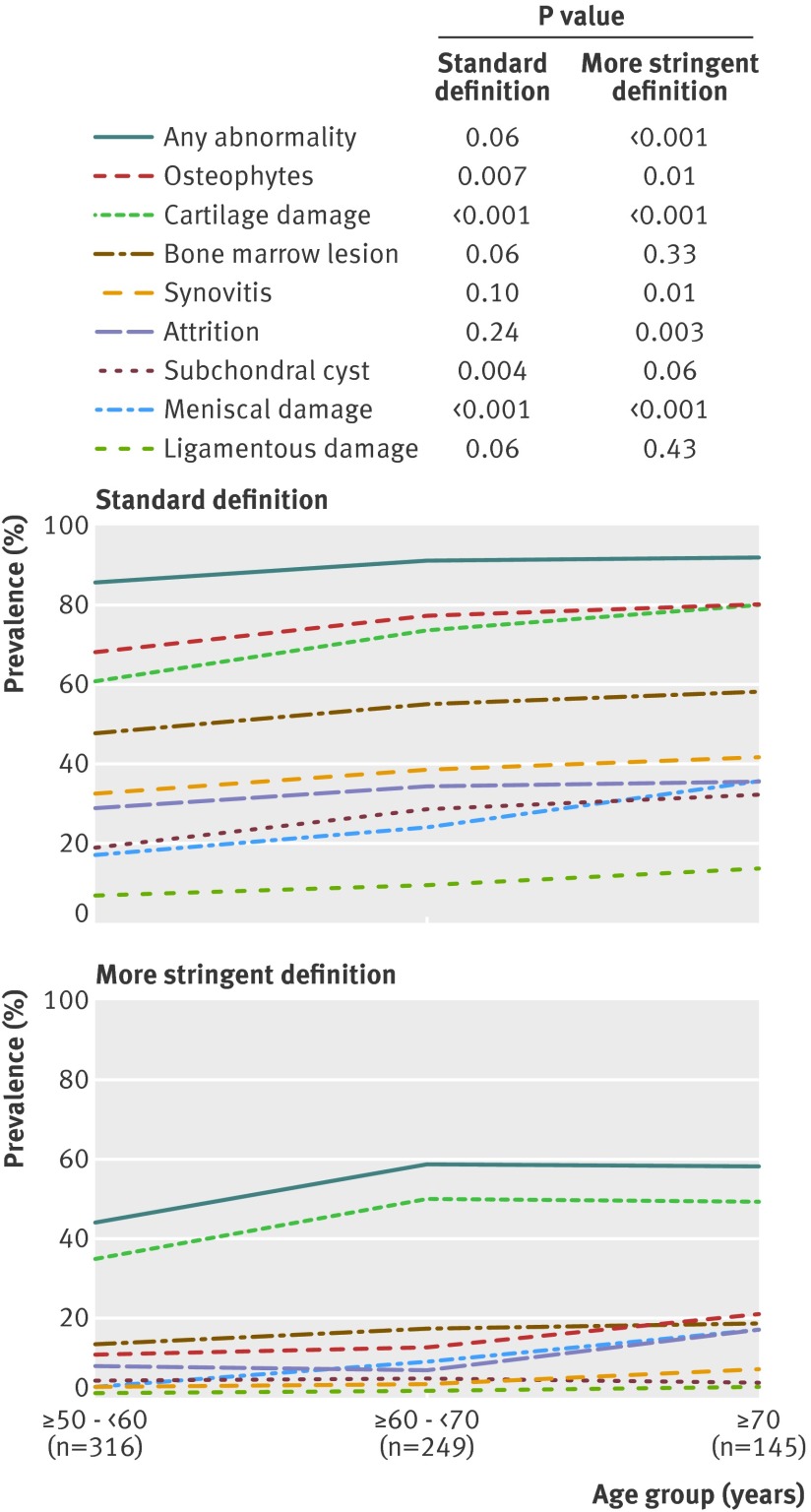
Results: Of the 710 participants, 393 (55%) were women, 660 (93%) were white, and 206 (29%) had knee pain in the past month. The mean age was 62.3 years and mean BMI was 27.9. Prevalence of "any abnormality" was 89% (631/710) overall. Osteophytes were the most common abnormality among all participants (74%, 524/710), followed by cartilage damage (69%, 492/710) and bone marrow lesions (52%, 371/710). The higher the age, the higher the prevalence of all types of abnormalities detectable by MRI. There were no significant differences in the prevalence of any of the features between BMI groups. The prevalence of at least one type of pathology ("any abnormality") was high in both painful (90-97%, depending on pain definition) and painless (86-88%) knees.
Conclusions: MRI shows lesions in the tibiofemoral joint in most middle aged and elderly people in whom knee radiographs do not show any features of osteoarthritis, regardless of pain.
Conflict of interest statement
Competing interests: All authors have completed the ICMJE uniform disclosure form at
Figures
References
-
- Sacks JJ, Luo YH, Helmick CG. Prevalence of specific types of arthritis and other rheumatic conditions in the ambulatory health care system in the United States, 2001-2005. Arthritis Care Res (Hoboken) 2010;62:460-4. - PubMed
-
- Bedson J, Jordan K, Croft P. The prevalence and history of knee osteoarthritis in general practice: a case-control study. Fam Pract 2005;22:103-8. - PubMed
-
- Felson DT, Zhang Y. An update on the epidemiology of knee and hip osteoarthritis with a view to prevention. Arthritis Rheum 1998;41:1343-55. - PubMed
Publication types
MeSH terms
Grants and funding
LinkOut - more resources
Full Text Sources
Medical
Miscellaneous