. 2012 Dec;61(12):3181-8.
doi: 10.2337/db12-0559. Epub 2012 Aug 28.

Altered MAPK signaling in progressive deterioration of endothelial function in diabetic mice

Affiliations

Altered MAPK signaling in progressive deterioration of endothelial function in diabetic mice

An Huang et al. Diabetes. 2012 Dec.

Abstract

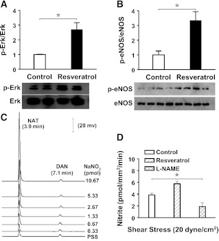
We aimed to investigate specific roles of mitogen-activated protein kinases (MAPK) in the deterioration of endothelial function during the progression of diabetes and the potential therapeutic effects of MAPK inhibitors and agonists in the amelioration of endothelial function. Protein expression and phosphorylation of p38, c-Jun NH(2)-terminal kinase (JNK), and extracellular signal-regulated kinase (Erk) were assessed in mesenteric arteries of 3- (3M) and 9-month-old (9M) male diabetic and control mice. The expression of p38, JNK, and Erk was comparable in all groups of mice, but the phosphorylation of p38 and JNK was increased in 3M and further increased in 9M diabetic mice, whereas the phosphorylation of Erk was substantially reduced in 9M diabetic mice. NADPH oxidase-dependent superoxide production was significantly increased in vessels of two ages of diabetic mice. Inhibition of either p38 with SB203580 or JNK with SP600125 reduced superoxide production and improved shear stress-induced dilation (SSID) in 3M, but not in 9M, diabetic mice. Treating the vessels of 9M diabetic mice with resveratrol increased Erk phosphorylation and shear stress-induced endothelial nitric oxide synthase (eNOS) phosphorylation and activity, but resveratrol alone did not improve SSID. Administration of resveratrol and SB203580 or resveratrol and SP600125 together significantly improved SSID in vessels of 9M diabetic mice. The improved response was prevented by U0126, an Erk inhibitor. Thus, p38/JNK-dependent increase in oxidative stress diminished nitric oxide-mediated dilation in vessels of 3M diabetic mice. Oxidative stress and impaired Erk-dependent activation of eNOS exacerbates endothelial dysfunction in the advanced stage of diabetes.


Figures

FIG. 1.
FIG. 1.
Protein expression of p38 (A), JNK (B), and Erk (C) in mesenteric arteries of 3M and 9M male Leprdb−/− and their heterozygous littermates (Leprdb+/−; a model of normal control mice). Summary data were obtained from 6 blots for each protein. The total expressions of p38, JNK, and Erk were normalized by β-actin. Densitometric ratios of phosphorylated and total protein expression of p38, JNK, and Erk were compared directly. Summary data were presented in comparison with the average expression in 3M Leprdb+/− group. *Significant difference between groups.
FIG. 2.
FIG. 2.
Shear stress (20 dynes/cm2)–induced dilation in mesenteric arteries of 3M and 9M Leprdb−/− and Leprdb+/− mice, in control and in the presence of SB203580 (A), a p38 inhibitor, or SP600125 (B), a JNK inhibitor, respectively. l-NAME was used in the group of 3M Leprdb−/− mice, in the presence of SB203580 or SP600125, to inhibit NO synthesis. n = 6–8 per group. *Significant difference between groups. PD, passive diameter.
FIG. 3.
FIG. 3.
Shear stress (20 dynes/cm2)– and NO (acidified NaNO2)-induced dilation in mesenteric arteries of 3M (A) and 9M (B) Leprdb−/− mice in the control condition (CTR) and in the presence of VAS2870 (VAS; a NADPH oxidase inhibitor), SB203580 (SB), and SP600125 (SP), respectively. n = 8 per group. *Significant difference between groups.
FIG. 4.
FIG. 4.
Superoxide production in mesenteric arteries of 3M and 9M Leprdb−/− and Leprdb+/− mice. A: Lucigenin chemiluminescence detection of superoxide in control and in the presence of SB203580 (SB), SP600125 (SP), or VAS2870 (VAS), respectively. n = 6–8 per group. B and C: Confocal image analyses of DHE staining in the endothelial cell (EC) and smooth muscle cell (SMC) layers of mesenteric arteries, respectively. n = 4 per group. Data were normalized by the mean of 3M Leprdb+/− group and expressed as relative fluorescent intensity of DHE staining. *Significant difference between groups.
FIG. 5.
FIG. 5.
Shear stress (20 dynes/cm2, 10 min)–stimulated Erk (A) and eNOS (B) phosphorylation (p) in mesenteric arteries of 9M Leprdb−/− mice in control and after treatment with resveratrol (100 mol/L) for 60 min. Data were summarized from 3 blots. *Significant difference between groups. C: HPLC/fluorescence detection of 1-(H)-naphthotriazole (NAT), a fluorescent product of nitrite and DAN. Traces show fluorescent signals and retention times of NAT and DAN, obtained from standard curves of sodium nitrite (0.33–10.67 pmol in 20 µL, equivalent to 20–640 μmol/L of sodium nitrite). D: Shear stress–induced release of NO (perfusate nitrite) in mesenteric arteries of 9M Leprdb−/− mice in the control condition and in the presence of resveratrol and resveratrol plus l-NAME. n = 8 per group. *Significant difference between groups.
FIG. 6.
FIG. 6.
Shear stress (20 dynes/cm2)–induced dilation in mesenteric arteries of 9M Leprdb−/− and Leprdb+/− mice, in control and in the presence of resveratrol. n = 6–8 per group. *Significant difference between groups. PD, passive diameter.
FIG. 7.
FIG. 7.
Shear stress (20 dynes/cm2)–induced dilation in mesenteric arteries of 9M Leprdb−/− mice in control and in the presence (+) or absence (─) of resveratrol, SB203580, SP600125, VAS2870, and U0126, respectively. n = 8 per group. *Significant difference between groups. PD, passive diameter.

Similar articles

Cited by

References

    1. Hamed S, Brenner B, Aharon A, Daoud D, Roguin A. Nitric oxide and superoxide dismutase modulate endothelial progenitor cell function in type 2 diabetes mellitus. Cardiovasc Diabetol 2009;8:56. - PMC - PubMed
    1. Hamed S, Brenner B, Abassi Z, Aharon A, Daoud D, Roguin A. Hyperglycemia and oxidized-LDL exert a deleterious effect on endothelial progenitor cell migration in type 2 diabetes mellitus. Thromb Res 2010;126:166–174 - PubMed
    1. Hamed S, Brenner B, Roguin A. Nitric oxide: a key factor behind the dysfunctionality of endothelial progenitor cells in diabetes mellitus type-2. Cardiovasc Res 2011;91:9–15 - PubMed
    1. Huang A, Yang YM, Feher A, Bagi Z, Kaley G, Sun D. Exacerbation of endothelial dysfunction during the progression of diabetes: role of oxidative stress. Am J Physiol Regul Integr Comp Physiol 2012;302:R674–R681 - PMC - PubMed
    1. Evans JL, Goldfine ID, Maddux BA, Grodsky GM. Oxidative stress and stress-activated signaling pathways: a unifying hypothesis of type 2 diabetes. Endocr Rev 2002;23:599–622 - PubMed

Publication types

MeSH terms