. 2012 Nov;86(22):12138-47.
doi: 10.1128/JVI.00375-12. Epub 2012 Aug 29.

Trafficking and replication patterns reveal splenic macrophages as major targets of dengue virus in mice

Affiliations

Trafficking and replication patterns reveal splenic macrophages as major targets of dengue virus in mice

Tyler R Prestwood et al. J Virol. 2012 Nov.

Abstract

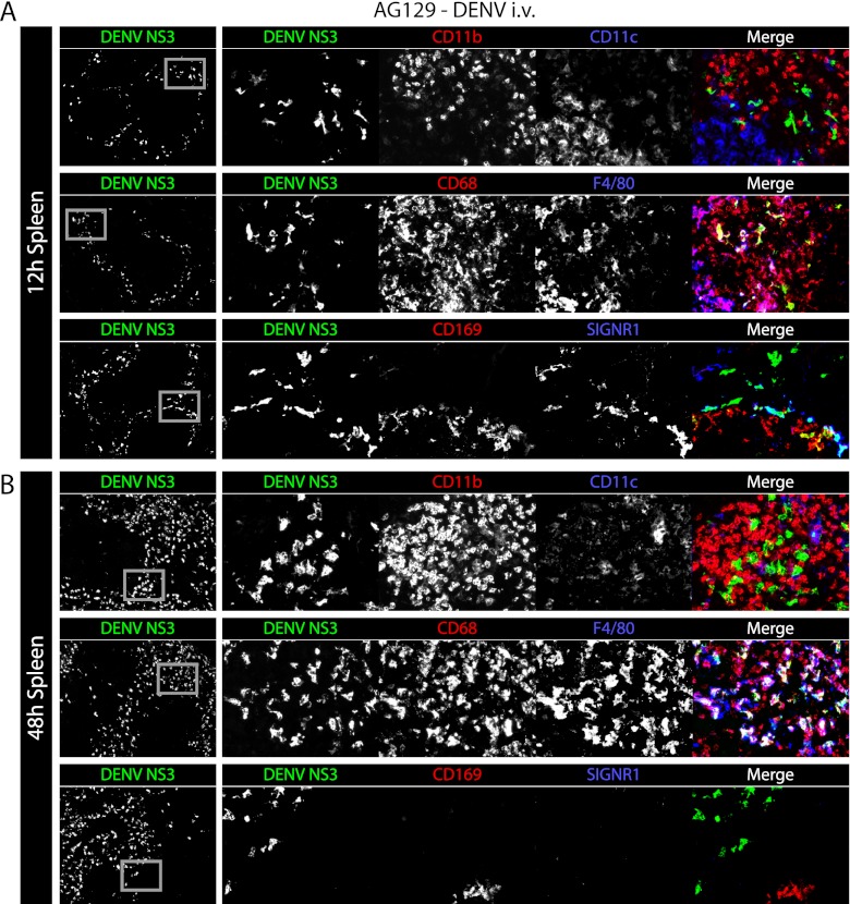
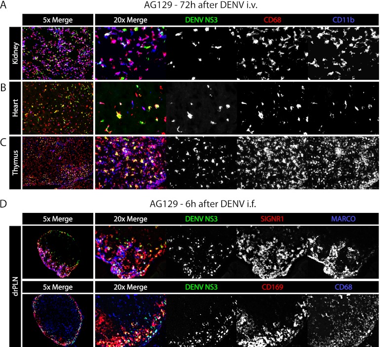
Human postmortem studies of natural dengue virus (DENV) infection have reported systemically distributed viral antigen. Although it is widely accepted that DENV infects mononuclear phagocytes, the sequence in which specific tissues and cell types are targeted remains uncharacterized. We previously reported that mice lacking alpha/beta and gamma interferon receptors permit high levels of DENV replication and show signs of systemic disease (T. R. Prestwood et al., J. Virol. 82:8411-8421, 2008). Here we demonstrate that within 6 h, DENV traffics to and replicates in both CD169(+) and SIGN-R1(+) macrophages of the splenic marginal zone or draining lymph node, respectively, following intravenous or intrafootpad inoculation. Subsequently, high levels of replication are detected in F4/80(+) splenic red pulp macrophages and in the bone marrow, lymph nodes, and Peyer's patches. Intravenously inoculated mice begin to succumb to dengue disease 72 h after infection, at which time viral replication occurs systemically, except in lymphoid tissues. In particular, high levels of replication occur in CD68(+) macrophages of the kidneys, heart, thymus, and gastrointestinal tract. Over the course of infection, proportionately large quantities of DENV traffic to the liver and spleen. However, late during infection, viral trafficking to the spleen decreases, while trafficking to the liver, thymus, and kidneys increases. The present study demonstrates that macrophage populations, initially in the spleen and other lymphoid tissues and later in nonlymphoid tissues, are major targets of DENV infection in vivo.


Figures

Fig 1
Fig 1
Dengue virus first infects the spleen, followed by bone marrow and then lymph nodes and Peyer's patches after intravenous inoculation. Representative immunohistochemical staining of tissue sections from naïve AG129 mice and AG129 mice at 6, 12, 24, 48, and 72 h after infection (1.5 × 10¹¹ GE of the DENV2 strain E124/128-IC i.v. on day 0). Spleens, cutaneous lymph nodes (CLNs: axillary, brachial, and inguinal lymph nodes), mesenteric lymph nodes (MLNs), Peyer's patches (PPs), and bone marrow (BM) were sectioned and stained for DENV NS3 (white when alone, green when shown with prM/M) and DENV prM/M (antibody clone 2H2, shown in red). The results are representative of 3 mice. WP labels the white pulp, LN labels the center of the lymph nodes, and PP labels the centers of the Peyer's patches.
Fig 2
Fig 2
Dengue virus infects systemic nonlymphoid tissues late during infection. Representative immunohistochemical staining of tissue sections from AG129 mice. (A) Tissues were harvested from naïve mice and mice 48 and 72 h after infection (1.5 × 10¹¹ GE of the DENV2 strain E124/128-IC i.v. on day 0) and stained for DENV NS3 (white) in the kidney, heart, thymus, adrenal gland, liver, lung, forestomach, stomach, duodenum, jejunum, ileum, cecum, colon, and rectum. The results are representative of 3 mice. (B) Tissues were harvested from mice 6, 24, and 72 h after infection (1.5 × 10¹¹ GE of the DENV2 strain E124/128-IC i.f. on day 0) and stained for DENV NS3 (white) in the spleen, draining popliteal lymph node (drPLN), contralateral popliteal lymph node (clPLN), cutaneous lymph nodes (CLNs: axillary, brachial, and inguinal lymph nodes), and kidney. The results are representative of 3 mice.
Fig 3
Fig 3
Schematic depicting the kinetics of DENV tissue tropism over the course of infection in mice. Model of DENV infection. DENV first infects the spleen followed by the bone marrow and then the lymph nodes and Peyer's patches (LN, PP). Infection decreases in these tissues while it increases systemically in nonlymphoid tissues, at which point mice become moribund.
Fig 4
Fig 4
Blood-borne DENV particles traffic mainly to the spleen and liver. (A) Representative immunohistochemical staining of spleen, liver, thymus, and kidney from naïve AG129 mice and from AG129 mice 6, 12, 18, 24, 36, 48, and 72 h after infection (1.5 × 10¹¹ GE of the DENV2 strain E124/128-IC i.v. on day 0 and 5 × 10¹⁰ GE DiD-labeled DENV2 strain E124/128-IC i.v. 2 h before harvest). Sections were stained for DENV NS3 (white, top row of each panel), and the DiD in the tissue was imaged (white, middle row of each panel). Merged images are shown on the bottom row of each panel (NS3 is shown as green, and DiD is shown as red). (B) Representative immunohistochemical staining of spleen from AG129 (top) or WT129 (bottom) mice 6 h after infection (5 × 10¹⁰ GE of DiD-labeled DENV2 strain E124/128-IC i.v.). Sections were stained for CD169, SIGN-R1, and MARCO (white, left), and the DiD in the tissue was imaged (white, middle). The merged image is shown on the right (DiD is shown as green, and CD169, SIGN-R1, and MARCO are shown as red). (C) Summary of results from panel A. The DiD intensity was quantified from 6 to 24 images per mouse with 5 mice per time point. Symbols and error bars represent the means ± SEMs. P values from two-tailed unpaired t tests: *, P ≤ 0.05; **, P ≤ 0.01; *** P ≤ 0.001.
Fig 5
Fig 5
In the spleen, DENV first infects macrophages of the marginal zone and later infects macrophages of the red pulp. (A) Representative immunohistochemical staining of spleen sections from AG129 mice 12 h after infection (5 × 10⁸ GE of the DENV2 strain E124/128-IC i.v. on day 0). Tissue sections were stained for DENV NS3 (green) and CD11b and CD11c (red and blue, respectively, top row), CD68 and F4/80 (red and blue, respectively, middle row), and CD169 and SIGN-R1 (red and blue, respectively, bottom row). Low-magnification (×5) images are shown on the left with DENV NS3 alone, while each individual channel and the merged image are shown on the right. Gray boxes on the DENV NS3 panels depict the areas of enlargement shown on the right. The results are representative of 3 mice. (B) Representative immunohistochemical staining of spleen sections from AG129 mice 48 h after infection (5 × 10⁸ GE of the DENV2 strain E124/128-IC i.v. on day 0). Sections are stained as described for panel A. The results are representative of 3 mice.
Fig 6
Fig 6
IFN-γ receptor signaling limits DENV replication in splenic red pulp macrophages. (A) Representative immunohistochemical staining of spleen sections from AG129 or A129 mice 12, 24, 48, and 72 after infection (5 × 10⁸ GE of the DENV2 strain E124/128-IC i.v. on day 0). Tissue sections were stained for F4/80 (red), DENV NS3 (green), and CD68 (blue). Low-magnification (×5) merged images are shown on the left as merged images only, medium-magnification (×20) images are shown in the middle as merged images only, and high-magnification (×33) images are shown on the right as the merged image followed by each individual channel (F4/80, DENV NS3, and CD68). Gray boxes on the left depict the area of enlargement shown on the immediate right. Two enlarged areas from each middle-magnification image are shown. The results are representative of 3 mice. (B) Representative immunohistochemical staining of spleen sections from AG129 or A129 mice 12, 24, 48, and 72 after infection (5 × 10⁸ GE of the DENV2 strain E124/128-IC i.v. on day 0), with each time point represented by a row. Images are arranged as described for panel A, except tissue sections were stained for CD169 (red), DENV NS3 (green), and SIGN-R1 (blue). The results are representative of 3 mice.
Fig 7
Fig 7
DENV infects macrophages of the kidney, heart, thymus, and lymph nodes. (A) Representative immunohistochemical staining of kidney, heart, and thymus sections from AG129 mice 72 h after infection (1.5 × 10¹¹ GE of the DENV2 strain E124/128-IC i.v. on day 0). Tissue sections were stained for DENV NS3 (green), CD68 (red), and CD11b (blue). On the left are ×5-magnification images of all channels, and on the right are ×20-magnification images with each channel shown separately in white. The results are representative of 3 mice. (B) Representative immunohistochemical staining of draining popliteal lymph node sections from AG129 mice 6 h after infection (1.5 × 10¹¹ GE of the DENV2 strain E124/128-IC i.f.). Tissue sections were stained for DENV NS3 (green), SIGN-R1 (red), and MARCO (blue) in the upper panel and DENV NS3 (green), CD169 (red), and CD68 (blue) in the lower panel. On the left are ×5-magnification images of all channels, and on the right are ×20-magnification images with each channel shown separately in white. The results are representative of 3 mice.

Similar articles

Cited by

References

    1. Ashour J, et al. 2010. Mouse STAT2 restricts early dengue virus replication. Cell Host Microbe 8:410–421 - PMC - PubMed
    1. Balsitis SJ, et al. 2009. Tropism of dengue virus in mice and humans defined by viral nonstructural protein 3-specific immunostaining. Am. J. Trop. Med. Hyg. 80:416–424 - PubMed
    1. Balsitis SJ, et al. 2010. Lethal antibody enhancement of dengue disease in mice is prevented by Fc modification. PLoS Pathog. 6:e1000790 doi:10.1371/journal.ppat.1000790 - DOI - PMC - PubMed
    1. Bente DA, et al. 2010. Pathogenesis and immune response of Crimean-Congo hemorrhagic fever virus in a STAT-1 knockout mouse model. J. Virol. 84:11089–11100 - PMC - PubMed
    1. Bhoopat L, et al. 1996. Immunohistochemical characterization of a new monoclonal antibody reactive with dengue virus-infected cells in frozen tissue using immunoperoxidase technique. Asian Pac. J. Allergy Immunol. 14:107–113 - PubMed

Publication types

MeSH terms

Substances