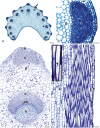
Collenchyma: a versatile mechanical tissue with dynamic cell walls
- PMID: 22933416
- PMCID: PMC3478049
- DOI: 10.1093/aob/mcs186
Collenchyma: a versatile mechanical tissue with dynamic cell walls
Abstract
Background: Collenchyma has remained in the shadow of commercially exploited mechanical tissues such as wood and fibres, and therefore has received little attention since it was first described. However, collenchyma is highly dynamic, especially compared with sclerenchyma. It is the main supporting tissue of growing organs with walls thickening during and after elongation. In older organs, collenchyma may become more rigid due to changes in cell wall composition or may undergo sclerification through lignification of newly deposited cell wall material. While much is known about the systematic and organographic distribution of collenchyma, there is rather less information regarding the molecular architecture and properties of its cell walls.
Scope and conclusions: This review summarizes several aspects that have not previously been extensively discussed including the origin of the term 'collenchyma' and the history of its typology. As the cell walls of collenchyma largely determine the dynamic characteristics of this tissue, I summarize the current state of knowledge regarding their structure and molecular composition. Unfortunately, to date, detailed studies specifically focusing on collenchyma cell walls have not been undertaken. However, generating a more detailed understanding of the structural and compositional modifications associated with the transition from plastic to elastic collenchyma cell wall properties is likely to provide significant insights into how specific configurations of cell wall polymers result in specific functional properties. This approach, focusing on architecture and functional properties, is likely to provide improved clarity on the controversial definition of collenchyma.
Figures


Similar articles
-
Comparative in situ analysis reveals the dynamic nature of sclerenchyma cell walls of the fern Asplenium rutifolium.Ann Bot. 2018 Feb 12;121(2):345-358. doi: 10.1093/aob/mcx167. Ann Bot. 2018. PMID: 29293865 Free PMC article.
-
Polysaccharide compositions of collenchyma cell walls from celery (Apium graveolens L.) petioles.BMC Plant Biol. 2017 Jun 15;17(1):104. doi: 10.1186/s12870-017-1046-y. BMC Plant Biol. 2017. PMID: 28619057 Free PMC article.
-
Developmental changes in collenchyma cell-wall polysaccharides in celery (Apium graveolens L.) petioles.BMC Plant Biol. 2019 Feb 19;19(1):81. doi: 10.1186/s12870-019-1648-7. BMC Plant Biol. 2019. PMID: 30782133 Free PMC article.
-
Effects of microgravity on the structure and function of plant cell walls.Int Rev Cytol. 1997;170:39-77. doi: 10.1016/s0074-7696(08)61620-4. Int Rev Cytol. 1997. PMID: 11536785 Review.
-
Feeding the Walls: How Does Nutrient Availability Regulate Cell Wall Composition?Int J Mol Sci. 2018 Sep 10;19(9):2691. doi: 10.3390/ijms19092691. Int J Mol Sci. 2018. PMID: 30201905 Free PMC article. Review.
Cited by
-
Ultrasound Pulse Emission Spectroscopy Method to Characterize Xylem Conduits in Plant Stems.Research (Wash D C). 2022 Sep 13;2022:9790438. doi: 10.34133/2022/9790438. eCollection 2022. Research (Wash D C). 2022. PMID: 36204251 Free PMC article.
-
Changes in the orientations of cellulose microfibrils during the development of collenchyma cell walls of celery (Apium graveolens L.).Planta. 2019 Dec;250(6):1819-1832. doi: 10.1007/s00425-019-03262-8. Epub 2019 Aug 28. Planta. 2019. PMID: 31463558
-
Phase-change-mediated transport and agglomeration of fungal spores on wheat awns.J R Soc Interface. 2022 May;19(190):20210872. doi: 10.1098/rsif.2021.0872. Epub 2022 May 18. J R Soc Interface. 2022. PMID: 35582813 Free PMC article.
-
Anatomical and Chemical Characterization of Ulmus Species from South Korea.Plants (Basel). 2021 Nov 29;10(12):2617. doi: 10.3390/plants10122617. Plants (Basel). 2021. PMID: 34961088 Free PMC article.
-
Ontogenetic tissue modification in Malus fruit peduncles: the role of sclereids.Ann Bot. 2014 Jan;113(1):105-18. doi: 10.1093/aob/mct262. Epub 2013 Nov 27. Ann Bot. 2014. PMID: 24287811 Free PMC article.
References
-
- Albersheim P, Darvill A, Roberts K, Sederoff R, Staehelin A. Plant cell walls: from chemistry to biology. New York: Garland Science; 2010.
-
- Alston HG. The subdivision of the Polypodiaceae. Taxon. 1956;5:23–25.
-
- von Alten H. Wurzelstudien. Botanische Zeitung. 1909;67:175–198.
-
- Ambronn H. Über die Entwickelungsgeschichte und die mechanishen Eigenschaften des Collenchyms. Jahrbuch für Wissenschaftliche Botanik. 1881;12:473–541.
-
- Anderson D. Über die Struktur der Kollenchymzellwand auf Grund mikrochemischer Untersuchungen. Sitzungsberichte der Akademie der Wissenschaften in Wien. Mathematisch-Naturwissenschaftliche Klasse. 1927;136:429–440.
Publication types
MeSH terms
LinkOut - more resources
Full Text Sources
Research Materials

