

Influence of cone beam CT on treatment plan before surgical intervention of mandibular third molars and impact of radiographic factors on deciding on coronectomy vs surgical removal
- PMID: 22933533
- PMCID: PMC5083118
- DOI: 10.1259/dmfr/98870341
Influence of cone beam CT on treatment plan before surgical intervention of mandibular third molars and impact of radiographic factors on deciding on coronectomy vs surgical removal
Abstract
Objectives: To assess the influence of cone beam CT (CBCT) on treatment plan before surgical intervention of mandibular third molars and to identify radiographic factors with an impact on deciding on coronectomy.
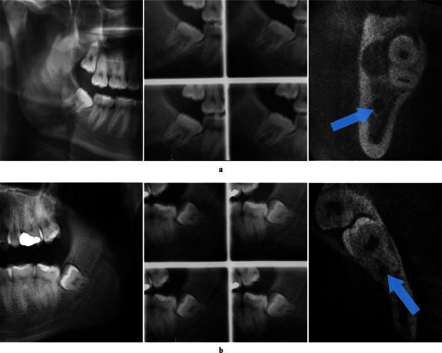
Methods: 186 mandibular third molars with an indication for surgical intervention underwent a radiographic examination with two methods: (1) panoramic imaging in combination with stereo-scanography and (2) CBCT. After the radiographic examination a treatment plan (TP) was established: either surgical removal (Sr) or coronectomy (Co). The first TP was based on the panoramic image and stereo-scanogram, while the second TP was established after CBCT was available. Logistic regression analyses were used to identify factors predisposing for Co after CBCT.
Results: Treatment was performed according to the second TP. Agreement between the first and second TP was seen in 164 cases (88%), while the TP changed for 22 teeth (12%) after CBCT. Direct contact between the third molar and the mandibular canal had the highest impact on deciding on Co [odds ratio (OR) = 101.8, p < 0.001]. Direct contact was not a sufficient factor, however; thus, lumen narrowing of the canal (OR = 38.9-147.2, p < 0.001) and canal positioned in a bending or a groove in the root complex (OR = 32.8, p = 0.016) were additional canal-related factors for deciding on Co.
Conclusion: CBCT influenced the treatment plan for 12%. Direct contact in combination with narrowing of the canal lumen and canal positioned in a bending or a groove in the root complex observed in CBCT images were significant factors for deciding on coronectomy.
Figures
References
-
- Renton T, Hankins M, Sproate C, McGurk M. A randomised controlled clinical trial to compare the incidence of injury to the inferior alveolar nerve as a result of coronectomy and removal of mandibular third molars. Br J Oral Maxillofac Surg 2005;43:7–12. - PubMed
-
- Leung YY, Cheung LK. Can coronectomy of wisdom teeth reduce the incidence of inferior dental nerve injury? Ann R Australas Coll Dent Surg 2008;19:50–51. - PubMed
-
- Cilasun U, Yildirim T, Guzeldemir E, Pektas ZO. Coronectomy in patients with high risk of inferior alveolar nerve injury diagnosed by computed tomography. J Oral Maxillofac Surg 2011;69:1557–1561. - PubMed
-
- Leung YY, Cheung LK. Safety of coronectomy versus excision of wisdom teeth: a randomized controlled trial. Oral Surg Oral Med Oral Pathol Oral Radiol Endod 2009;108:821–827. - PubMed
-
- Dolanmaz D, Yildirim G, Isik K, Kucuk K, Ozturk A. A preferable technique for protecting the inferior alveolar nerve: coronectomy. J Oral Maxillofac Surg 2009;67:1234–1238. - PubMed
Publication types
MeSH terms
LinkOut - more resources
Full Text Sources
Other Literature Sources
Research Materials
