Review
. 2012 Sep;76(3):497-529.
doi: 10.1128/MMBR.00006-12.

The role of bacterial enhancer binding proteins as specialized activators of σ54-dependent transcription

Affiliations
Review

The role of bacterial enhancer binding proteins as specialized activators of σ54-dependent transcription

Matthew Bush et al. Microbiol Mol Biol Rev. 2012 Sep.

Abstract

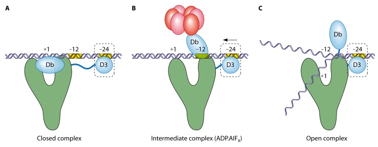
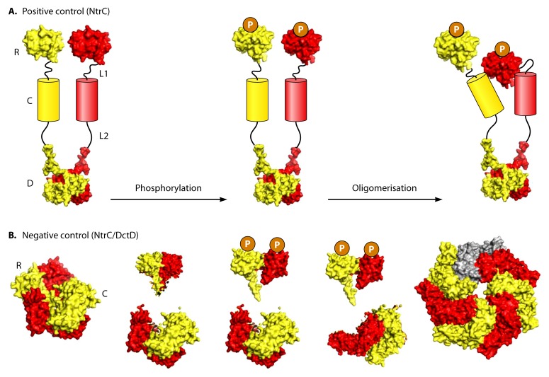
Bacterial enhancer binding proteins (bEBPs) are transcriptional activators that assemble as hexameric rings in their active forms and utilize ATP hydrolysis to remodel the conformation of RNA polymerase containing the alternative sigma factor σ(54). We present a comprehensive and detailed summary of recent advances in our understanding of how these specialized molecular machines function. The review is structured by introducing each of the three domains in turn: the central catalytic domain, the N-terminal regulatory domain, and the C-terminal DNA binding domain. The role of the central catalytic domain is presented with particular reference to (i) oligomerization, (ii) ATP hydrolysis, and (iii) the key GAFTGA motif that contacts σ(54) for remodeling. Each of these functions forms a potential target of the signal-sensing N-terminal regulatory domain, which can act either positively or negatively to control the activation of σ(54)-dependent transcription. Finally, we focus on the DNA binding function of the C-terminal domain and the enhancer sites to which it binds. Particular attention is paid to the importance of σ(54) to the bacterial cell and its unique role in regulating transcription.


Figures

Fig 1
Fig 1
Initiation of transcription by the RNAP-σ70 (A) and RNAP-σ54 (B) holoenzymes. The σ70 factor directs the binding of polymerase to the consensus −10 (TATAAT) and −35 (TTGACA) sequences to form an energetically unfavorable closed complex (CC) that is readily converted into an open complex (OC) to initiate transcription. In contrast, the σ54 factor directs the binding of RNAP to conserved −12 (TGC) and −24 (GG) promoter elements that are part of the wider consensus sequence YTGGCACGrNNNTTGCW (where uppercase type indicates highly conserved residues, lowercase type indicates weakly conserved residues, N is nonconserved, Y is pyrimidines, R is purines, and W is A or T) (10). This forms an energetically favorable CC that rarely isomerizes into the OC. In order to form the transcription “bubble,” a specialized activator (a bacterial enhancer binding protein [bEBP]) must bind and use the energy from ATP hydrolysis to remodel the holoenzyme.
Fig 2
Fig 2
Activation of bacterial transcription initiation (σ54 family dependent). (A) σ54 directs the RNA polymerase holoenzyme to bind at the −12 and −24 promoter elements. The interaction of the bacterial enhancer binding protein (bEBP) with the σ54-RNAP holoenzyme is dependent on the binding of the activator (shown as an oligomer in green) to upstream activator sequences (UASs), 80 to 150 bp upstream of the transcriptional start site. (B) DNA looping occurs, often facilitated by other proteins such as integration host factor (IHF), enabling bEBP-σ54 interactions. In σ54-dependent transcription, the closed complex does not spontaneously undergo isomerization (melting of the double-stranded DNA). (C) Nucleotide hydrolysis by the activator promotes remodeling of the closed complex through a series of protein-protein and protein-DNA interactions that promote the formation of the open complex (30).
Fig 3
Fig 3
Domain organization of σ54. E. coli σ54 (residues 1 to 477) consists of 3 regions (regions I to III). DNA binding motifs include the DNA cross-linking (X link) region, the helix-turn-helix (HTH) motif, and the RpoN box, all present at the C terminus. Region I interacts with the activator of transcription. Region II is often acidic and occasionally absent. The location of the main core RNAP binding determinants (residues 120 to 215) is shown (30).
Fig 4
Fig 4
Schematic representation of the proposed relative positions and movements of σ54 domains and promoter DNA in the closed (A), intermediate (B), and open (C) complexes. The DNA binding region (D3 density) and region I (Db density) of σ54 are shown in light blue, with the core enzyme in green. The bEBP/activator is shown as a hexamer in red, and the promoter DNA is shown in dark blue. Cryo-EM structures of the RNAP-σ54 holoenzyme indicate that the “Db density” is in close proximity to the −12 position of the promoter DNA. Therefore, it has been proposed that region I of σ54 prevents the initiation of transcription by obstructing the loading of DNA into the active-site channel of the core RNAP. The activator (i) causes the melting of DNA at the −12 position, (ii) interacts with region I to relocate the Db density, and (iii) results in the downstream movement of the DNA binding (D3) region of σ54, bringing the origin of DNA melting (−12) near the active site. (Adapted from reference with permission from Elsevier.)
Fig 5
Fig 5
Domain architecture of the five classical groups (groups I to V) of bEBPs (218). The central AAA+ domain (C) (red) is highly conserved and absolutely essential for σ54-dependent transcription. The C-terminal DNA binding domain (D) (green) consists of an HTH motif that directs the bEBP to specific UAS/enhancer binding sites and is absent in some bEBPs (group V). The N-terminal regulatory domain (R) is not well conserved between members of the bEBP family. Different sensory domains are present depending on the environmental signal to be detected, but in some bEBPs, they are absent (group IV). Group I bEBPs contain a response regulator (RR) domain (blue). Group II bEBPs contain Per, ARNT, and Sim (PAS) domains (orange) or XylR-N and V4R (vinyl 4 reductase) domains (pink). Group III bEBPs contain a cGMP-specific and stimulated phosphodiesterase, Anabaena adenylate cyclase, and E. coli FhlA (GAF) domain (purple). HrpR and HrpS are coactivators of transcription and therefore are grouped together.
Fig 6
Fig 6
Domain map and sequence alignment of the conserved regions of bEBP AAA+ domains (C1 to C7) (148). The conserved regions are based on a structure-based sequence alignment (186). Key residues (Walker A, “switch” Asn, GAFTGA, Walker B, and R fingers) are highlighted in yellow, and nonconsensus sequences in the alignments are highlighted in red. The locations of loop 1, loop 2, sensor I, and sensor II motifs are indicated with their sequences highlighted in gray. Alignments were conducted by using ClustalW (www.ebi.ac.uk/clustalw/), using the following sequences from UniProtKB/Swiss-Prot (http://www.expasy.ch/): PspF (E. coli), NifA (A. vinelandii), XylR (P. putida), DmpR (Pseudomonas sp.), NtrC (E. coli), ZraR (E. coli), NtrC1 (A. aeolicus), NtrC4 (A. aeolicus), FlgR (H. pylori), DctD (S. meliloti), FhlA (E. coli), HrpR (P. syringae), NorR (E. coli), and TyrR (E. coli). The R (regulatory) and D (DNA binding) domains are also illustrated, although they are not to scale.
Fig 7
Fig 7
Crystal structure of the ATP-bound NtrC1 E239A variant (PDB accession number 3M0E) (51). (A) Close-up of the nucleotide hydrolysis site in NtrC1 in the ATP-bound state that forms at the interface between two adjacent protomers in the bEBP oligomer. The Walker A (WA) (GxxxxGK) residues are labeled in brown, the Walker B (hhhhDE) residues are labeled in cyan, sensor I (SI) is shown in magenta, the conserved “switch” asparagine (N195) is shown in yellow, sensor II is shown in red, and the putative in trans R fingers (R162 and R168) are shown in green. The location of ATP and its γ-phosphate is also indicated. Walker A forms a P loop that interacts with the phosphates of ATP. The Walker B aspartate has a role in the coordination of Mg2+ (shown as a pink sphere), and the glutamate residue is thought to activate a water molecule (shown in blue) for the nucleophilic attack of the γ-phosphate. The conserved asparagine functions in the hydrolysis-dependent “switch” (173). The sensor I threonine residue (T279) has been implicated in the coupling of nucleotide hydrolysis to conformational change (173). Sensor II residues are located in the third helix of the α-helical subdomain. The R fingers have been implicated in intersubunit catalysis and nucleotide sensing (92, 133, 155). (B) The structure of the NtrC1 E239A variant is composed of 7 monomers (alternate light and dark shading) arranged in a ring (seen from below). The conserved elements in the bEBP subfamily are highlighted as described above for panel A, with the key surface-exposed loop 1 (L1) in light blue, the highly conserved GAFTGA motif emphasized in dark blue, and side chains displayed. Nucleotide-dependent conformational changes in L1 are assisted via movements of a second loop, loop 2 (L2), which is shown in orange. (C) Each bEBP monomer is made up of an α/β subdomain followed by a smaller α-helical subdomain. The hydrolysis site is found at the cleft between these subdomains and between adjacent protomers. Key motifs are colored as described above for panels A and B.
Fig 8
Fig 8
(A and B) Structure of monomeric PspF1–275 bound to ATP (PDB accession number 2C96/2C9C) (A) and ADP (PDB accession number 2C98/2C9C) (B). Important motifs are highlighted: Walker A (brown), Walker B residue E108 (cyan), sensor I residue T148 (magenta), and “switch” residue N64 (yellow). Loop 1 (L1) and loop 2 (L2) are labeled, with the nonresolved fold of L1 indicated with a red dotted line. (C) Switching mechanism of the Walker B E108 residue. In the ATP-bound state, E108 interacts with N64 (indicated by a black dotted line). In the ADP-bound state, there is a 90° rotation in the N64 side chain so that E108 interacts with sensor I residue T148 via a water molecule (indicated by a red dotted line).
Fig 9
Fig 9
Summary of the nucleotide-driven conformational changes that occur during ATP hydrolysis, as proposed for PspF (173). The ground (blue), transition (red), posthydrolysis (green), and released (purple) states are indicated. For simplicity, only the “switch” interactions are shown with the associated relocations of linker 1, helix 3 (H3), and L1/L2. (Adapted from reference with permission from Elsevier.)
Fig 10
Fig 10
In cis and in trans interactions predicted to form during nucleotide hydrolysis in PspF. Interactions in cis center around the E97 (green) residue, which interacts with either R131 (blue) (ATP state) or R91 (purple) (ADP state), depending on the position of the Walker B-asparagine “switch.” Upon γ-phosphate release, E97 breaks its interaction with R131, allowing R131 to instead contact R81 (red). These new interactions result in the compaction of L1 and L2 down toward the surface of the AAA+ domain, enabling σ54 relocation. Interactions in trans center around the E130 residue (orange) (subunit n), which contacts the R98 residue (magenta) (subunit n+1) in the ATP-bound state but which interacts with the R95 residue (cyan) (subunit n+1) in the ADP-bound state. (A) Region of the crystal structure of PspF1–275 in the ATP-bound form (PDB accession number 2C96) and the ADP-bound form (PDB accession number 2C98) showing the locations of the key residues involved in inter- and intrasubunit interactions. Interactions occurring within the protomer are indicated by double-headed arrows. (B) Schematic showing the in cis and in trans interactions that occur before and after γ-phosphate release. Interactions are indicated by arrows, and residue colors correspond to those described above for panel A. (Adapted from reference with permission of the publisher.)
Fig 11
Fig 11
Comparison of the ADP-bound and ATP-bound structures of the AAA+ domains of NtrC1 and the NtrC1 E339A variant, respectively. In each structure, the proposed R fingers are indicated (R293 and R299) in dark green. The sensor II arginine (R357) is shown in red. The Walker B “DE” residues (D238 and E239) are shown in cyan. The E242 (orange) and E256 (blue) residues may form interprotomer interactions. The asparagine “switch” proposed for PspF is shown in yellow (N195 in NtrC1). The K250 residue (magenta) exists in two distinct conformations depending on whether ATP or ADP is bound; this residue is proposed to be important for mediating the transition of loop 1 (L1) and loop 2 (L2) to a raised conformation. (Left) The ADP-bound structure of the NtrC1 AAA+ domain (PDB accession number 1NY6 [B-C interface] [128]). (Right) The ATP-bound structure of the NtrC1(E239A) AAA+ domain (PDB accession number 3MOE [51]). The magnesium ion is shown as a light pink sphere, and the active water molecule is shown as a blue sphere.
Fig 12
Fig 12
Models for the coordination of nucleotide hydrolysis between protomers in the AAA+ hexamer. In solution, the majority of bEBPs exist in equilibrium between dimeric and hexameric forms. In the case of PspF, binding of ATP or ADP promotes hexamerization and ATP hydrolysis. (A) The stochastic model assumes that each catalytic site is independent. (D) The synchronized/concerted model is based on homogeneous nucleotide occupancy and assumes that each site simultaneously hydrolyzes ATP. (B and C) Data for PspF suggest that ATP hydrolysis occurs via either the rotational (B) or sequential (C) mechanisms that utilize heterogeneous nucleotide occupancy. Both models are based on cooperativity between protomers in the hexamer. (Adapted from reference with permission of the publisher.)
Fig 13
Fig 13
Negative (A) and positive (B) control of AAA+ domain activity. In the more common mechanism of negative control, ligand binding (or phosphorylation) relieves the repression of the regulatory (R) domain on the central (C) domain, which is intrinsically competent to hydrolyze ATP. The AAA+ domain is then able to carry out ATP hydrolysis. Accordingly, when the R domain is removed, the bEBP is active irrespective of the presence or absence of a signaling molecule (shown as a purple triangle) or an available kinase. In positive control, ligand binding or phosphorylation has a genuine stimulatory function. The phosphorylated or ligand-bound form of the R domain activates the C domain, which is not intrinsically competent to hydrolyze ATP. The AAA+ domain is then able to carry out ATP hydrolysis. Accordingly, when the R domain is removed, the bEBP is inactive irrespective of the presence or absence of a signaling molecule or an available kinase.
Fig 14
Fig 14
Possible targets of regulatory domain-mediated regulation. (A) Most commonly, the regulatory domain represses oligomerization, e.g., NtrC1 and DctD (66, 128), or promotes self-association in response to the signal, e.g., NtrC (62). (B) In PspF, negative regulation directly targets the nucleotide hydrolysis machinery (115). While the binding of ATP releases L1 and L2 to establish a weak interaction with σ54, hydrolysis is required to produce a strong interaction that results in the remodeling of the holoenzyme (173). (C) NorR may represent a newly identified mechanism of control in which the interaction with σ54 is the target of the regulatory domain. Where oligomerization is not the target, the preassembly of a hexamer prior to activation may have a physiological advantage, e.g., a rapid response to stress. The mechanisms of regulation that target ATP hydrolysis, oligomerization, and σ54 interactions are likely to be highly interconnected, with the enzymatic activity of the AAA+ domain being the ultimate target of regulation.
Fig 15
Fig 15
Models of bEBP activation by phosphorylation through the promotion of oligomerization by stimulatory (A) and derepressing (B) functions of the response regulator (RR) domain. In activated NtrC, the DNA binding domain is hidden underneath the hexamer ring. For DctD and NtrC1, no information is available to define the positions of DNA binding domains. R, regulatory domain; L1, linker 1; C, central domain; L2, linker 2; D, DNA binding domain. Models were built by using published structures of NtrC fragments R (off state, PDB accession number 1KRW; on state, accession number 1KRX) and L2-D (PDB accession number 1NTC) and NtrC1 fragments R (PDB accession number 1ZY2), R-L1-C (PDB accession number 1NY5), and L1-C (PDB accession number 1NY6). (Adapted from reference with permission from Elsevier.)
Fig 16
Fig 16
Negative regulation of PspF AAA+ activity by PspA targets the nucleotide hydrolysis machinery via the W56 residue. (A) Crystal structure of PspF1–275 (PDB accession number 2C96) in the ATP-bound state showing the key residues involved. (B) Model of the signaling pathway coupling negative regulation to substrate remodeling. PspA interacts directly with PspF, an interaction that is detected via the surface-exposed W56 (purple) residue of PspF. W56 relays this information to the conserved asparagine (N64) (red) via β-sheet 2 (blue). This causes the repositioning of the Walker B glutamate (E108) (green) to prevent ATP hydrolysis. Upon the dissipation of the PMF, PspA inhibition is prevented (possibly facilitated by PspB and PspC), and ATP hydrolysis can occur, strengthening the σ54 interaction and leading to substrate remodeling. The removal of the γ-phosphate leads to a 90° rotation of the E108 side chain, breaking the interaction with N64. This change is translated to GAFTGA-containing L1 (orange) via helix 3 (H3) (yellow), and the loops compact back downwards. (Adapted from reference with permission of the publisher.)
Fig 17
Fig 17
Model of NorR-dependent activation of norVW (adapted from reference 37). (A) The binding of NorR to the norR-norVW intergenic region that contains the three NorR binding sites (sites 1, 2, and 3) (highlighted in red) is thought to facilitate the formation of a higher-order oligomer that is most likely to be a hexamer (207). (B) Although bound to DNA, in the absence of NO, the N-terminal GAF domains (blue rectangles) negatively regulate the activity of the AAA+ domains (green circles) by preventing the access of the surface-exposed loops to σ54. (C) In the “on” state, NO binds to the iron center in the GAF domain, forming a mononitrosyl iron species. The repression of the AAA+ domain is relieved, enabling ATP hydrolysis by NorR coupled to conformational changes in the AAA+ domain. (D) In the presence of ATP, the surface-exposed loops (orange) that include the GAFTGA motifs move into an extended conformation to establish an initial interaction with σ54 that is strengthened upon hydrolysis, resulting in the remodeling of the closed complex. Upon phosphate release, L1 and L2 compact downwards, enabling the relocation of the sigma factor (173). For simplicity, DNA is not illustrated in panel B, C, or D.
Fig 18
Fig 18
Schematic representation of σ54 remodeling by NtrC. Conformational changes in the GAFTGA loops of the central (C) domain (red and yellow circles) and the DNA binding (D) domain (blue and purple ovals) in activated NtrC are shown. The regulatory (R) domains (red and yellow squares) are shown in the phosphorylated form that promotes hexamerization. NtrC dimers are thought to bind to two UAS sequences and, once activated, recruit a further dimer from solution in order to oligomerize. The D domains are located on the bottom of the bEBP ring, whereas the GAFTGA loops contact σ54 (orange) from the top. In the transition state of ATP hydrolysis, the interaction between the GAFTGA loops and σ54 is strengthened, and the DNA binding domains form a tight association with the oligomeric ring. Upon phosphate release, the loops disengage σ54, and the tight constraints upon enhancer DNA are relaxed. (Adapted from reference with permission of the publisher.)
None
None

Similar articles

Cited by

References

    1. Adams H, Teertstra W, Demmers J, Boesten R, Tommassen J. 2003. Interactions between phage-shock proteins in Escherichia coli. J. Bacteriol. 185:1174–1180 - PMC - PubMed
    1. Ades SE. 2006. AAA+ molecular machines: firing on all cylinders. Curr. Biol. 16:R46–R48 - PubMed
    1. Akoev V, Gogol EP, Barnett ME, Zolkiewski M. 2004. Nucleotide-induced switch in oligomerization of the AAA+ ATPase ClpB. Protein Sci. 13:567–574 - PMC - PubMed
    1. Alfano JR, Collmer A. 1997. The type III (Hrp) secretion pathway of plant pathogenic bacteria: trafficking harpins, Avr proteins, and death. J. Bacteriol. 179:5655–5662 - PMC - PubMed
    1. Aravind L, Ponting CP. 1997. The GAF domain: an evolutionary link between diverse phototransducing proteins. Trends Biochem. Sci. 22:458–459 - PubMed

Publication types

MeSH terms

LinkOut - more resources