Review
. 2012 Sep;76(3):565-84.
doi: 10.1128/MMBR.00014-12.

Regulation of the histidine utilization (hut) system in bacteria

Affiliations
Review

Regulation of the histidine utilization (hut) system in bacteria

Robert A Bender. Microbiol Mol Biol Rev. 2012 Sep.

Abstract

The ability to degrade the amino acid histidine to ammonia, glutamate, and a one-carbon compound (formate or formamide) is a property that is widely distributed among bacteria. The four or five enzymatic steps of the pathway are highly conserved, and the chemistry of the reactions displays several unusual features, including the rearrangement of a portion of the histidase polypeptide chain to yield an unusual imidazole structure at the active site and the use of a tightly bound NAD molecule as an electrophile rather than a redox-active element in urocanase. Given the importance of this amino acid, it is not surprising that the degradation of histidine is tightly regulated. The study of that regulation led to three central paradigms in bacterial regulation: catabolite repression by glucose and other carbon sources, nitrogen regulation and two-component regulators in general, and autoregulation of bacterial regulators. This review focuses on three groups of organisms for which studies are most complete: the enteric bacteria, for which the regulation is best understood; the pseudomonads, for which the chemistry is best characterized; and Bacillus subtilis, for which the regulatory mechanisms are very different from those of the Gram-negative bacteria. The Hut pathway is fundamentally a catabolic pathway that allows cells to use histidine as a source of carbon, energy, and nitrogen, but other roles for the pathway are also considered briefly here.


Figures

Fig 1
Fig 1
The two conserved histidine utilization (Hut) pathways. Pathway 1 yields 1 mole each of ammonia, glutamate, and formamide per mole of histidine. Pathway 2 yields 2 moles of ammonia, 1 mole of glutamate, and 1 mole of formate per mole of histidine.
Fig 2
Fig 2
Formation of MIO, an unusual imidazole structure generated at the active site of histidase, as described by Schwede et al. (143).
Fig 3
Fig 3
Urocanase reaction. Hydration of the Cformula imageC double bond of urocanate generates hydroxyimidazole propionate, which spontaneously undergoes an enol-keto tautomerization to imidazolone propionate (IP).
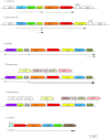
Fig 4
Fig 4
Genetic structure of the hut operons. Genes encoding elements of the Hut pathway are shown on the main line and in bold colors. Genes with the same name and same color are homologous. [Note that hutG of the enteric bacteria is not homologous to hutG(Ps) of the pseudomonads.] Genes of unknown function that are not part of the hut pathway are listed with orf names rather than hut names and appear on lines above the main line. orfH (sometimes called hutH1) is similar to hutH, but its gene product lacks histidase activity. orfT is similar to hutT, but its gene product cannot replace the hutT gene product for transporting histidine or urocanate. orfA, -B, -C, -D, -H, and -T may have functions that are related to histidine degradation (see the text) and are shown in pastel colors. Genes shown without color are unrelated to hut. Arrows under the main line indicate transcription units and directions. The illustration was drawn approximately to scale.
Fig 5
Fig 5
Regulatory sites in the hutU promoter region of K. pneumoniae. The upper line represents the intergenic region between hutC and hutU, with +1 indicating the start of transcription of the hutUHT operon. Boxes represent matches to consensus sequences for known regulators of hutUHT transcription, and the lines beneath the boxes represent the extents of the footprints of those regulators on the DNA. The diagram was drawn approximately to scale.
Fig 6
Fig 6
Regulatory sites in the control region of the hut operon of B. subtilis.The upper line represents the control region of the hutHUIGM operon, including the start of transcription (+1) and the hutP gene. Boxes below that line indicate the binding sites for the following regulatory proteins: CodY, a transcriptional repressor that binds to the hutOA site; CcpA, a protein that blocks transcription by binding at a cre site; and HutP, which prevents transcriptional termination at the hutC site, indicated by the lollipop structure on the upper line. The diagram was drawn approximately to scale.
None

References

    1. Akashi H, Gojobori T. 2002. Metabolic efficiency and amino acid composition in the proteomes of Escherichia coli and Bacillus subtilis. Proc. Natl. Acad. Sci. U. S. A. 99:3695–3700 - PMC - PubMed
    1. Allison SL, Phillips AT. 1990. Nucleotide sequence of the gene encoding the repressor for the histidine utilization genes of Pseudomonas putida. J. Bacteriol. 172:5470–5476 - PMC - PubMed
    1. Ames GF. 1964. Uptake of amino acids by Salmonella typhimurium. Arch. Biochem. Biophys. 104:1–18 - PubMed
    1. Atkinson MR, Wray LVJ, Fisher SH. 1990. Regulation of histidine and proline degradation enzymes by amino acid availability in Bacillus subtilis. J. Bacteriol. 172:4758–4765 - PMC - PubMed
    1. Baldauf SL, Cardani MA, Bender RA. 1988. Regulation of the galactose-inducible lac operon and the histidine utilization operons in pts mutants of Klebsiella aerogenes. J. Bacteriol. 170:5588–5593 - PMC - PubMed

MeSH terms

LinkOut - more resources