. 2012 Aug 29;32(35):11919-29.
doi: 10.1523/JNEUROSCI.3852-11.2012.

Adaptor protein APPL1 couples synaptic NMDA receptor with neuronal prosurvival phosphatidylinositol 3-kinase/Akt pathway

Affiliations

Adaptor protein APPL1 couples synaptic NMDA receptor with neuronal prosurvival phosphatidylinositol 3-kinase/Akt pathway

Yu-bin Wang et al. J Neurosci. .

Abstract

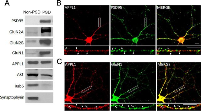
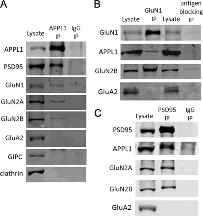
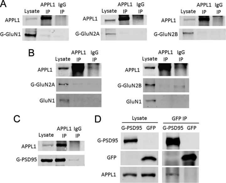
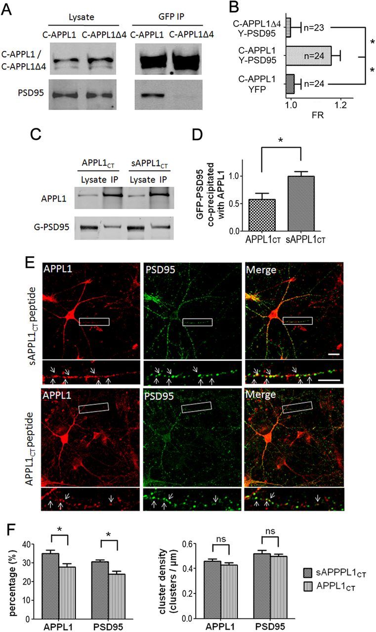
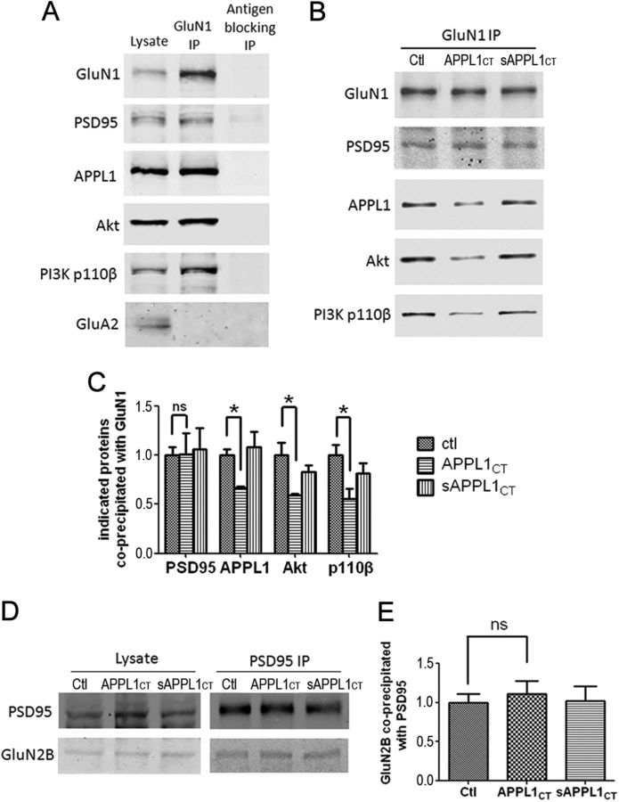
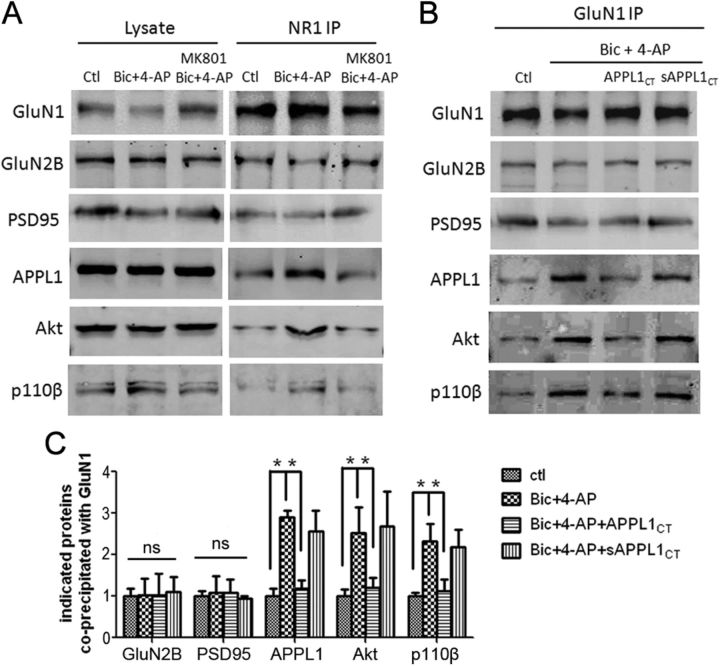
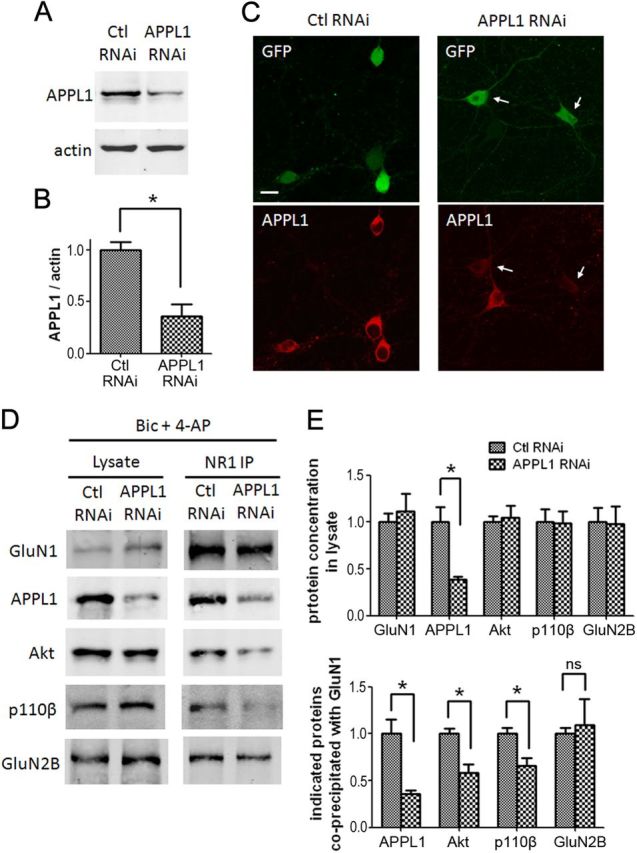
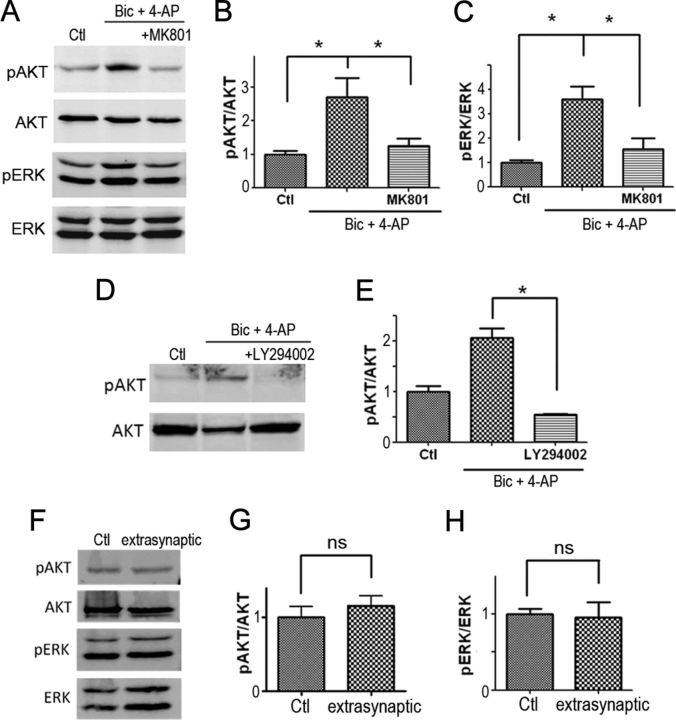
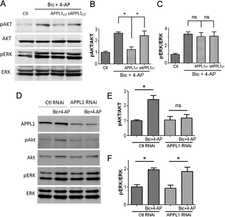
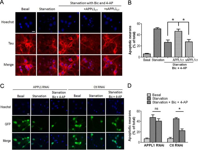
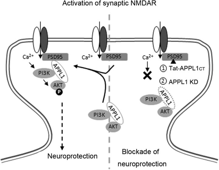
It is well known that NMDA receptors (NMDARs) can both induce neurotoxicity and promote neuronal survival under different circumstances. Recent studies show that such paradoxical responses are related to the receptor location: the former to the extrasynaptic and the latter to the synaptic. The phosphoinositide 3-kinase (PI3K)/Akt kinase cascade is a key pathway responsible for the synaptic NMDAR-dependent neuroprotection. However, it is still unknown how synaptic NMDARs are coupled with the PI3K/Akt pathway. Here, we explored the role of an adaptor protein-adaptor protein containing pH domain, PTB domain, and leucine zipper motif (APPL1)-in this signal coupling using rat cortical neurons. We found that APPL1 existed in postsynaptic densities and associated with the NMDAR complex through binding to PSD95 at its C-terminal PDZ-binding motif. NMDARs, APPL1, and the PI3K/Akt cascade formed a complex in rat cortical neurons. Synaptic NMDAR activity increased the association of this complex, induced activation of the PI3K/Akt pathway, and consequently protected neurons against starvation-induced apoptosis. Perturbing APPL1 interaction with PSD95 by a peptide comprising the APPL1 C-terminal PDZ-binding motif dissociated the PI3K/Akt pathway from NMDARs. Either the peptide or lentiviral knockdown of APPL1 blocked synaptic NMDAR-dependent recruitment and activation of PI3K/Akt pathway, and consequently blocked synaptic NMDAR-dependent neuroprotection. These results suggest that APPL1 contributes to connecting synaptic NMDARs with the intracellular PI3K/Akt cascade and the downstream prosurvival signaling pathway in rat cortical neurons.


Figures

Figure 1.
Figure 1.
Subcellular localization of APPL1 protein in brain. A, APPL1 and Akt existed both in the non-PSD and PSD fractions. PSD95, GluN2A, GluN2B, and GluN1 are components of the PSD fraction. Rab5 is a component of the late endosome. Synaptophysin is a component of presynaptic structure. B, Cultured cortical neurons (DIV 11–13) coimmunostained for endogenous APPL1 and PSD95. APPL1 clusters partially colocalized with PSD95 clusters (arrows). Scale bar, 10 μm. C, Coimmunostaining of endogenous APPL1 and GluN1. APPL1 clusters partially colocalized with GluN1 clusters (arrows). Scale bar, 10 μm.
Figure 2.
Figure 2.
APPL1 associates with the NMDA receptor complex in brain. A, APPL1 coprecipitated PSD95, GluN1, GluN2A, and GluN2B, but did not coprecipitate GluA2, GIPC, and clathrin from lysate of mouse cortex. Immunoprecipitation with nonimmune rabbit IgG was included as a negative control. B, GluN1 coprecipitated APPL1 and GluN2B but not GluA2 from lysate of mouse cortex. Immunoprecipitation with GluN1 antibody preblocked by excess antigen peptide (antigen blocking) was included as a negative control. C, PSD95 coprecipitated APPL1, GluN2A, and GluN2B, but not GluA2 from lysate of mouse cortex.
Figure 3.
Figure 3.
APPL1 interacts with PSD95 but not with NMDAR subunits. A, Co-IP of transfected GFP-GluN1, GFP-GluN2A, or GFP-GluN2B with endogenous APPL1 in HEK293 cells. GFP-GluN1, GFP-GluN2A, or GFP-GluN2B did not coprecipitate with APPL1. B, GFP-GluN2A/GluN1 or GFP-GluN2B/GluN1 were cotransfected into HEK293 so that they could form functional NMDARs on the plasma membrane. APPL1 was still unable to coprecipitate GluN1, GluN2A, or GluN2B under such conditions. C, GFP-PSD95 was transfected into HEK293 cells. APPL1 coprecipitated GFP-PSD95. D, GFP-PSD95 or GFP was transfected into HEK293 cells. GFP-PSD95 but not GFP coprecipitate APPL1.
Figure 4.
Figure 4.
APPL1 interacts with PSD95 through its C-terminal PDZ-binding motif. A, CFP-APPL1/PSD95 or CFP-APPL1Δ4/PSD95 was cotransfected into HEK293. CFP-APPL1 but not CFP-APPL1Δ4 coprecipitated PSD95. B, Three-cube FRET measurements in HEK293 cells. Coexpression of CFP-APPL1 and YFP-PSD95 yielded positive FRET signals while CFP-APPL1 and YFP, CFP-APPL1Δ4, and YFP-PSD95 did not. The horizontal axis indicates FRET ratio (FR). *p < 0.01, two-tailed t test. C, Application of 100 μm Tat-APPL1CT peptide (APPL1CT) on GFP-PSD95 transfected HEK293 cells for 2 h before cell harvest perturbed the co-IP of GFP-PSD95 with APPL1, compared with that of Tat-sAPPL1CT peptide (sAPPL1CT). D, Statistical analysis of C. Western results of three independent co-IP tests were analyzed. The ratio of the band intensity of coprecipitated GFP-PSD95 to precipitated APPL1 was compared between the APPL1CT group and the sAPPL1CT group. All ratios were normalized (divided by the average ratio of sAPPL1CT group) before t test (*p < 0.05, two-tailed t test; n = 3). E, Application of 10 μm Tat-APPL1CT peptide for 1 h on cultured cortical neurons perturbed the colocalizaion of endogenous APPL1 and PSD95, compared with that of Tat-sAPPL1CT peptide. Scale bar, 10 μm. F, Statistical analysis of E. Left, Tat-APPL1CT significantly decreased the percentage of APPL1 clusters that colocalized with PSD95 clusters and the percentage of PSD95 that colocalized with APPL1 (∼1000 clusters of each protein from three separate experiments; *p < 0.05, two-tailed t test). Right, Density of APPL1 or PSD95 clusters (clusters per micrometer of dendrite branch) was not affected by Tat-APPL1CT peptide (ns, no significant difference; two-tailed t test; n = 3). Error bars indicate SEM.
Figure 5.
Figure 5.
Disruption of APPL1 and NMDAR complex interaction dissociates PI3K and Akt from NMDARs. A, GluN1 coprecipitated PSD95, APPL1, Akt, and p110β but not GluA2 from lysate of cultured cortical neurons. Immunoprecipitation with GluN1 antibody preblocked by excess antigen peptide (antigen blocking) was included as the negative control. B, Incubation of cultured cortical neurons with Tat-APPL1CT peptide for 2 h partially blocked the co-IP of APPL1, Akt, and p110β with GluN1, but not the co-IP of PSD95 with GluN1. Treatment with Tat-sAPPL1CT peptide did not have the blocking effect. C, Statistical analysis of three independent co-IP tests. The ratio of the band intensity of coprecipitated PSD95, APPL1, Akt, or p110β to precipitated GluN1 was compared. All ratios were normalized (divided by the average ratio of control group) before t test (*p < 0.05; ns, no significant difference; two-tailed t test; n = 3). D, Co-IP of GluN2B with PSD95 was not perturbed by Tat-APPL1CT peptide. E, Statistical analysis of D (ns, no significant difference; two-tailed t test; n = 3). Error bars indicate SEM.
Figure 6.
Figure 6.
Synaptic NMDAR activity increases the association of APPL1/PI3K/Akt to the NMDAR complex. A, Treatment of cultured cortical neurons with 50 μm Bic and 250 μm 4-AP for 30 min increased the co-IP of APPL1, Akt, and p110β with GluN1, but not the co-IP of GluN2B or PSD95 with GluN1. MK801 (50 μm) blocked the Bic- and 4-AP-induced increase of interaction. B, Preincubation of cortical neurons with Tat-APPL1CT peptide for 1 h blocked the Bic- and 4-AP-induced increase of interaction. Tat-sAPPL1 CT peptide did not have the blocking effect. C, Statistical analysis of three independent co-IP tests. ns, No significant difference by one-way ANOVA (GluN2B, F(3,8) = 0.04, n = 3, p > 0.05; PSD95, F(3,8) = 0.15, n = 3, p > 0.05). *p < 0.05 by Bonferroni's multiple-comparisons test after significant one-way ANOVA (APPL1, F(3,8) = 31.55, n = 3, p < 0.05; Akt, F(3,8) = 7.62, n = 3, p < 0.05; p110β, F(3,8) = 8.30, n = 3, p < 0.05). Error bars indicate SEM.
Figure 7.
Figure 7.
Lentiviral RNAi of APPL1 blocks synaptic NMDAR-induced recruitment of Akt and PI3K to NMDARs. A, Cultured cortical neurons (DIV 6) were infected by APPL1 RNAi or control RNAi lentivirus. Six days after infection, neurons were harvested for Western blot. B, Quantification of blots from three independent experiments (*p < 0.05; two-tailed t test; n = 3). C, Immunofluorescent staining of infected neurons by APPL1 antibody. Neurons expressing GFP fluorescent indicate virus expression. The arrows indicate APPL1 knockdown neurons. Scale bar, 10 μm. D, Six days after lentiviral infection, neurons were treated for 30 min with Bic and 4-AP and processed for co-IP. E, Statistical analysis of three independent co-IP. Top, APPL1 knocking down did not affect the concentration of GluN1, Akt, p110β, and GluN2B in lysate. Bottom, APPL1 knocking down blocked the co-IP of APPL1, Akt, and p110β with GluN1, but did not affect the co-IP of GluN2B with GluN1 (*p < 0.05; ns, no significant difference; two-tailed t test; n = 3). Error bars indicate SEM.
Figure 8.
Figure 8.
Synaptic NMDARs but not extrasynaptic NMDARs induce Akt and ERK activation. A, Cultured cortical neurons (DIV 12–14) were treated for 30 min with 50 μm Bic and 250 μm 4-AP. MK801 (50 μm) was used to test whether the activation was NMDAR dependent. The levels of phospho-Akt Ser473 (pAkt), Akt, phospho-ERK1/2 (pErk), and ERK1/2 (Erk) were detected by Western blot. B, The ratio of phospho-Akt to Akt was compared between three groups. All ratios were normalized (divided by the average ratio of control group) before one-way ANOVA. *p < 0.05 by Bonferroni's multiple-comparisons test after significant one-way ANOVA (F(2,11) = 6.40; n = 5; p < 0.05). C, The ratio of phospho-ERK1/2 to ERK1/2 was compared between three groups. *p < 0.05 by Bonferroni's test after significant one-way ANOVA (F(2,6) = 11.50; n = 3; p < 0.05). D, Pretreatment with the PI3K inhibitor LY294002 (50 μm) for 5 min blocked the increase of phospho-Akt by Bic plus 4-AP treatment. E, Statistical analysis of D (*p < 0.05; two-tailed t test; n = 3). F, Extrasynaptic NMDARs were selectively activated by NMDA (10 μm), following irreversible blockade of synaptic NMDARs. G, H, Statistical analysis of F. Extrasynaptic stimulation did not increase pAkt/Akt or pERK/ERK (ns, no significant difference; two-tailed t test; n = 3). Error bars indicate SEM.
Figure 9.
Figure 9.
APPL1 is necessary for synaptic NMDAR-mediated AKT activation. A–C, Tat-APPL1CT peptide specifically blocked synaptic NMDAR-mediated AKT activation but not ERK1/2 activation. A, Cultured cortical neurons were preincubated with 10 μm Tat-APPL1CT or Tat-sAPPL1CT peptide for 1 h before treatment with Bic and 4-AP. The levels of pAkt, Akt, pErk, and Erk were detected by Western blot. B, Statistical analysis of A. Tat-APPL1CT, but not Tat-sAPPL1CT blocked the increase of phospho-Akt by Bic and 4-AP treatment. *p < 0.05 by Bonferroni's test after significant one-way ANOVA (F(3,8) = 11.68; n = 3; p < 0.05). C, Tat-APPL1CT or Tat-sAPPL1CT did not affect the increase of phospho-ERK1/2 by Bic and 4-AP treatment. ns, p > 0.05 by Bonferroni's test after significant one-way ANOVA (F(3,8) = 8.38; n = 3; p < 0.05). D–F, Lentiviral RNAi of APPL1 specifically blocked synaptic NMDAR-mediated AKT activation but not ERK1/2 activation. D, Lentivirus-infected neurons were treated for 30 min with Bic and 4-AP and processed for Western blot. E, Statistical analysis of D. APPL1 RNAi blocked Akt activation induced by Bic and 4-AP, while control RNAi did not (*p < 0.05; ns, no significant difference; two-tailed t test; n = 3). F, Neither APPL1 RNAi nor control RNAi affected ERK1/2 activation induced by Bic and 4-AP (*p < 0.05; two-tailed t test; n = 3). Error bars indicate SEM.
Figure 10.
Figure 10.
APPL1 is required for synaptic NMDA receptor-dependent neuroprotection. A, Cultured cortical neurons (DIV 10) were subjected to trophic deprivation (starvation) for 3 d. Incubation of starved neurons with 50 μm Bic and 250 μm 4-AP protected them from apoptosis. Tat-APPL1CT or Tat-sAPPL1CT peptide was applied at 10 μm along with Bic and 4-AP to test whether it blocked the neuroprotective effect of Bic and 4-AP. Hoechst staining was then used to quantify the death rate of neurons. Hoechst brightly stains the condensed or fragmented nuclei of apoptotic neurons and more dimly stains the normal nuclei of healthy neurons. Tau protein was simultaneously immunostained to reveal the morphology of neurons. Note that the Tau staining of Hoechst-positive neurons was unclear, suggesting the unhealthy state of these neurons. Scale bar, 10 μm. B, Quantification data of A. Tat-APPL1CT but not Tat-sAPPL1CT increased the proportion of apoptotic neurons, suggesting that Tat-APPL1CT blocked the neuroprotective effect of Bic and 4-AP. *p < 0.05 by Bonferroni's test after significant one-way ANOVA (F(4,10) = 28.78; n = 3; p < 0.05); 300–500 cells for each group in one independent experiment. C, Neurons at DIV 6 were infected by APPL1 RNAi or control RNAi lentivirus. Five days after infection, neurons were subjected to trophic deprivation for another 3 d, with or without the addition of Bic and 4-AP. The proportion of apoptotic neurons was analyzed by Hoechst staining. Neurons expressing GFP fluorescence indicate virus expression. D, Quantification data of C. APPL1 RNAi blocked the neuroprotective effect of Bic and 4-AP, while control RNAi did not (*p < 0.05; ns, no significant difference; two-tailed t test; n = 3; 300–500 cells for each group in one independent experiment). Error bars indicate SEM.
Figure 11.
Figure 11.
Schematic illustration of the involvement of APPL1 in synaptic NMDA receptor-dependent neuroprotection. Left, Synaptic NMDAR activity induces the recruitment of APPL1/PI3K/Akt to the NMDAR complex. APPL1 binds to the NMDAR complex through the PDZ interaction with PSD95. The formation of this complex facilitates the signaling from NMDARs to the Akt pathway. Local Ca2+ influx via NMDARs activates calmodulin. Calmodulin directly binds to and activates PI3K (Joyal et al., 1997) or indirectly activates PI3K through the Ras pathway (Sutton and Chandler, 2002). PI3K then phosphorylates and activates Akt, and phospho-Akt phosphorylates and regulates many intracellular substrates implicated in neuroprotection. Right, Treatment with Tat-APPL1CT peptide that blocks the APPL1–PSD95 interaction (1) or knockdown of APPL1 (2) dissociates PI3K and Akt from the NMDAR complex. Under this circumstance, synaptic NMDAR activity does not activate the PI3K/Akt cascade or the downstream prosurvival pathway.

Similar articles

Cited by

References

    1. Aarts M, Iihara K, Wei WL, Xiong ZG, Arundine M, Cerwinski W, MacDonald JF, Tymianski M. A key role for TRPM7 channels in anoxic neuronal death. Cell. 2003;115:863–877. - PubMed
    1. Alessi DR, James SR, Downes CP, Holmes AB, Gaffney PR, Reese CB, Cohen P. Characterization of a 3-phosphoinositide-dependent protein kinase which phosphorylates and activates protein kinase Balpha. Curr Biol. 1997;7:261–269. - PubMed
    1. Cleasby ME, Lau Q, Polkinghorne E, Patel SA, Leslie SJ, Turner N, Cooney GJ, Xu A, Kraegen EW. The adaptor protein APPL1 increases glycogen accumulation in rat skeletal muscle through activation of the PI3-kinase signalling pathway. J Endocrinol. 2011;210:81–92. - PMC - PubMed
    1. Downward J. How BAD phosphorylation is good for survival. Nat Cell Biol. 1999;1:E33–E35. - PubMed
    1. Hardingham GE. Coupling of the NMDA receptor to neuroprotective and neurodestructive events. Biochem Soc Trans. 2009;37:1147–1160. - PMC - PubMed

Publication types

MeSH terms

Substances

LinkOut - more resources