. 2012 Aug 29;32(35):11942-55.
doi: 10.1523/JNEUROSCI.1330-12.2012.

The C-type natriuretic peptide induces thermal hyperalgesia through a noncanonical Gβγ-dependent modulation of TRPV1 channel

Affiliations

The C-type natriuretic peptide induces thermal hyperalgesia through a noncanonical Gβγ-dependent modulation of TRPV1 channel

Lipin Loo et al. J Neurosci. .

Abstract

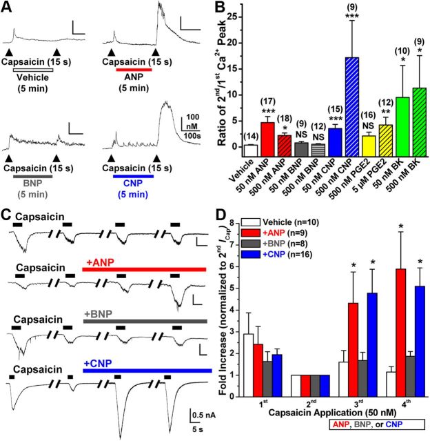
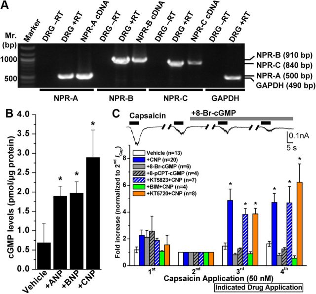
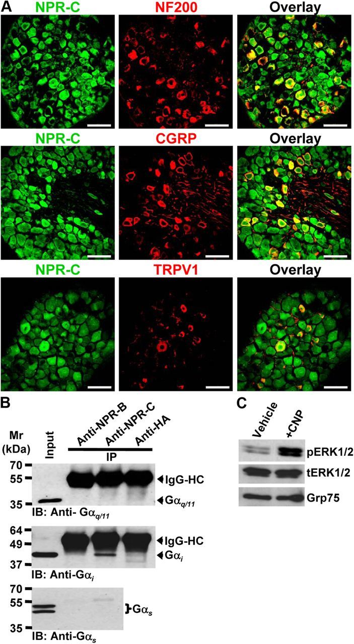
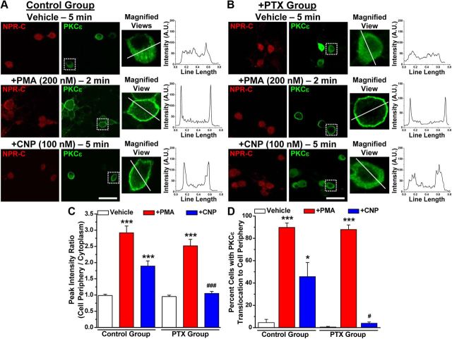
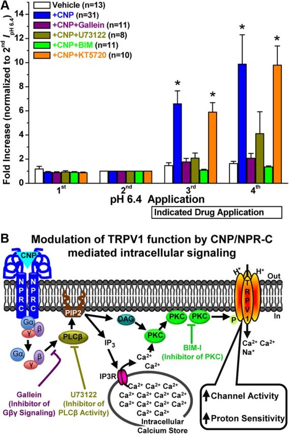
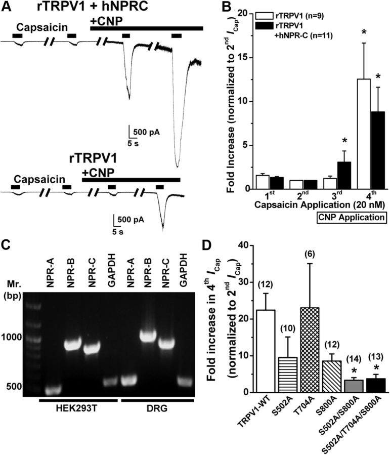
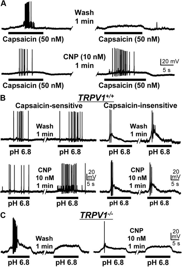
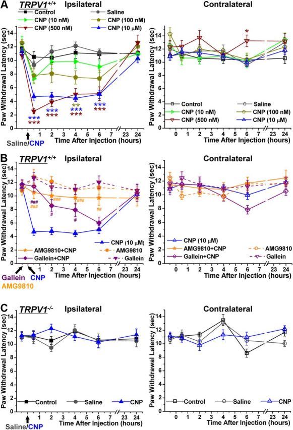
Natriuretic peptides (NPs) control natriuresis and normalize changes in blood pressure. Recent studies suggest that NPs are also involved in the regulation of pain sensitivity, although the underlying mechanisms remain essentially unknown. Many biological effects of NPs are mediated by guanylate cyclase (GC)-coupled NP receptors, NPR-A and NPR-B, whereas the third NP receptor, NPR-C, lacks the GC kinase domain and acts as the NP clearance receptor. In addition, NPR-C can couple to specific Gα(i)-Gβγ-mediated intracellular signaling cascades in numerous cell types. We found that NPR-C is coexpressed in transient receptor potential vanilloid-1 (TRPV1)-expressing mouse dorsal root ganglia (DRG) neurons. NPR-C can be coimmunoprecipitated with Gα(i), and C-type natriuretic peptide (CNP) treatment induced translocation of protein kinase Cε (PKCε) to the plasma membrane of these neurons, which was inhibited by pertussis toxin pretreatment. Application of CNP potentiated capsaicin- and proton-activated TRPV1 currents in cultured mouse DRG neurons and increased their firing frequency, an effect that was absent in DRG neurons from TRPV1(-/-) mice. CNP-induced sensitization of TRPV1 activity was attenuated by pretreatment of DRG neurons with the specific inhibitors of Gβγ, phospholipase C-β (PLCβ), or PKC, but not of protein kinase A, and was abolished by mutations at two PKC phosphorylation sites in TRPV1. Furthermore, CNP injection into mouse hindpaw led to the development of thermal hyperalgesia that was attenuated by administration of specific inhibitors of Gβγ or TRPV1 and was also absent in TRPV1(-/-) mice. Thus, our work identifies the Gβγ-PLCβ-PKC-dependent potentiation of TRPV1 as a novel signaling cascade recruited by CNP in mouse DRG neurons that can lead to enhanced nociceptor excitability and thermal hypersensitivity.


Figures

Figure 1.
Figure 1.
Natriuretic peptides ANP and CNP, but not BNP, sensitize TRPV1 channel activity in mouse DRG neurons. A, Representative traces of Ca2+ flux in small/medium-diameter cultured mouse DRG neurons in response to two successive applications of capsaicin (50 nm, 15 s, arrowheads), with the continuous extracellular perfusion of ANP, BNP, and CNP (50 nm each) or vehicle for 5 min between two capsaicin applications. B, Quantification of Ca2+ flux measurements from Ca2+ imaging experiments as shown in A, with extracellular perfusion of 50 and 500 nm ANP, BNP, CNP, or BK, and 500 nm or 5 μm PGE2. Data are presented as mean ± SEM of the ratio of second versus first Ca2+ peak; n values are shown in parentheses for each treatment group. *p < 0.05, **p < 0.01, and ***p < 0.001, significantly different (NS, not significant) compared with vehicle (Kruskal–Wallis test with Dunn's correction). C, Representative traces of whole-cell TRPV1 currents in small/medium-diameter cultured mouse DRG neurons in response to four successive applications of capsaicin (50 nm for 5 s; with 1 min interval), with continuous extracellular perfusion of ANP, BNP, or CNP (10 nm) or vehicle (extracellular buffer) after the second capsaicin application. D, Quantification of capsaicin-activated TRPV1 currents (ICap) for experiments as shown in C. Data are presented as mean ± SEM of fold increase in peak ICap normalized to second ICap; n values are given for each treatment group. *p < 0.05, significantly different compared with the respective ICap for vehicle group (one-way ANOVA with post hoc Dunnett's correction).
Figure 2.
Figure 2.
Expression of NP receptors and role of downstream cGMP signaling in mouse DRG neurons underlying CNP modulation of TRPV1 channel activity. A, All three NP receptors (NPR-A, NPR-B, and NPR-C) are expressed in DRG neurons. Representative RT-PCR analysis of NPR expression in cultured mouse DRG neurons with primers specific for NPR-A, NPR-B, and NPR-C, as well as for GAPDH as the housekeeping gene. B, ELISA-based quantification of intracellular cGMP levels in isolated mouse DRG neurons after ANP, BNP, or CNP treatment (100 nm for 30 min). Data are presented as mean ± SEM (n = 3). *p < 0.05, significantly different compared with the vehicle group (Student's t test). C, CNP-induced sensitization of TRPV1 is dependent on PKC, but not cGMP–PKG or PKA, signaling. Top panel shows representative traces of TRPV1 currents in small/medium-diameter cultured mouse DRG neurons in response to four successive applications of capsaicin (50 nm for 5 s; with 1 min interval), with continuous extracellular perfusion of 8-Br-cGMP (100 μm) after the second Icap. Bottom panel shows the quantification of ICap with indicated drug treatments [100 μm 8-Br-cGMP, 200 μm 8-pCPT-cGMP, 10 nm CNP, 10 nm CNP + 500 nm KT5823 (PKG inhibitor), 10 nm CNP + 400 nm KT5720 (PKA inhibitor), and 10 nm CNP + 1 μm BIM (PKC inhibitor)]. Peak ICap amplitudes are normalized to second ICap application of the respective vehicle or treatment groups, and the data are presented as mean ± SEM of fold-increase in ICap compared with second ICap (respective n values are shown in the figure). *p < 0.05, significantly different compared with the vehicle group (one-way ANOVA with post hoc Dunnett's correction).
Figure 3.
Figure 3.
Expression of NPR-C and characterization of downstream intracellular signaling originating from CNP treatment in mouse DRG neurons. A, Representative photomicrographs depicting immunohistochemical staining of mouse L4–L6 DRG sections, indicating NPR-C expression in NF200-positive large-diameter neurons, as well as in CGRP- and TRPV1-positive small/medium-diameter neurons. Scale bar, 50 μm. B, Immunoprecipitation of Gαi subunits, but not Gαq or Gαs subunits, with anti-NPR-C antibody, but not with anti-NPR-B antibody, in cultured mouse DRG neurons. Anti-HA antibody was used as a negative control in immunoprecipitation reactions. The immunoprecipitates, along with the input DRG lysates, were size fractionated in SDS-PAGE and immunoblotted with specific antibodies against Gαi, Gαq, and Gαs subunits. C, Western/immunoblot analysis of ERK1/2 phosphorylation induced by CNP treatment of cultured mouse DRG neurons (100 nm for 30 min). Blots were probed with phospho-ERK1/2 and total ERK1/2, as well as with GRP75 as loading control. IB, Immunoblot; IP, immunoprecipitation.
Figure 4.
Figure 4.
CNP treatment leads to translocation of PKCε in mouse DRG neurons. Immunocytochemical analysis of PKCε translocation to the cell periphery in cultured mouse DRG neurons upon treatment with CNP (100 nm for 5 min) and PMA (200 nm for 2 min; as a positive control) compared with vehicle (DMSO) treatment. Representative photomicrographs of neurons immunostained with anti-NPR-C (red) and anti-PKCε (green) antibodies under control (A) and PTX (100 μm, 24 h) (B) treatment conditions. Right panels for each image sets are magnified views of cells marked with respective rectangular white boxes, and the line graphs show the PKCε distribution profile (NIH Image J) within respective cells across the drawn white lines. The high-intensity peak signals near both ends of the path described by the line denote increased PKCε translocation to the cell plasma membrane. C, Quantification of PKCε translocation to the cell periphery under different treatment conditions (for details of analysis, see Materials and Methods). Data are presented at mean ± SEM of the peak intensity ratios (peripheral/cytoplasmic; n = 25 cells for each treatment condition, from four independent cultures). D, Quantification of number of DRG neurons with PKCε translocation to the cell periphery under different treatment conditions. Data are presented at mean ± SEM of the percentage of cells with increased PKCε staining intensity at the cell periphery (n > 500 neurons for each treatment group, from 4 independent cultures). For C, D, *p < 0.05 and ***p < 0.001, significantly different compared with their respective vehicle groups; #p < 0.05 and ###p < 0.001, significantly different compared with CNP treatment in control group (one-way ANOVA with post hoc Bonferroni's correction).
Figure 5.
Figure 5.
CNP specifically sensitizes TRPV1-mediated acidic pH-induced inward currents in mouse DRG neurons. A, Representative sets of traces of four successive proton (pH 6.4 extracellular buffer for 5 s; with 1 min interval) activated currents (IpH 6.4) in capsaicin-sensitive (left panel sets) and capsaicin-insensitive (right panel sets) small/medium-diameter cultured mouse DRG neurons, with or without continuous extracellular perfusion of CNP (10 nm) after the second IpH 6.4. B, Representative sets of traces of four successive IpH 6.4 recorded from small/medium-diameter cultured mouse DRG neurons obtained from TRPV1−/− mice, with or without continuous extracellular perfusion of CNP (10 nm) after the second IpH 6.4. C, Quantification of IpH 6.4 from experiments as shown in A and B. Data are presented as mean ± SEM of fold increase in peak IpH 6.4 normalized to the second IpH 6.4, and n values are shown in parentheses for each genotype/current/treatment group. *p < 0.05, significantly different compared with the respective IpH 6.4 for the vehicle group (one-way ANOVA with post hoc Dunnett's correction). D, CNP treatment (10 nm for 5 min) led to a leftward shift in the pH dose–response relationship of IpH in capsaicin-sensitive small/medium-diameter cultured mouse DRG neurons. Data are presented as mean ± SEM of inward currents in response to pH 7.4, 6.8, 6.4, 6.8, 5.4, and 4.8, normalized to the peak amplitude of IpH 4.8, and fitted with the Hill equation. The n values and EC50 pH values are mentioned in the panel for vehicle and CNP treatment groups.
Figure 6.
Figure 6.
Gβγ/PLCβ/PKC-mediated intracellular signaling underlies CNP-induced sensitization of TRPV1 in mouse DRG neurons. A, Quantified data are shown from whole-cell voltage-clamp experiments with four successive IpH 6.4, with continuous extracellular application of CNP (10 nm), with or without pharmacological modulators after the second IpH 6.4 in capsaicin-sensitive small/medium-diameter cultured mouse DRG neurons, as in experiments shown in Figure 5, A and C. Data for vehicle and CNP treatment groups from Figure 4C are replotted here for comparison. All the data are presented as mean ± SEM of fold increase in peak IpH 6.4 normalized to the second IpH 6.4, and n values are given for each treatment group. *p < 0.05, significantly different compared with the respective IpH 6.4 for the vehicle group (one-way ANOVA with post hoc Dunnett's correction). The CNP-induced potentiation of IpH 6.4 was significantly reduced by pretreatment with the inhibitors of Gβγ-mediated activation of PLCβ (gallein, 100 μm), PLCβ activity (U73122, 5 μm), and PKC activity (BIM, 1 μm), but not by the inhibitor of PKA activity (KT5720, 400 nm). B, A schematic model of CNP-induced sensitization of TRPV1 channel activity via Gβγ–PLCβ–PKC signaling in mouse DRG neurons, as verified experimentally in A.
Figure 7.
Figure 7.
CNP/NPR-C/PKC-induced sensitization of TRPV1 activity is dependent on two PKC phosphorylation sites in the channel protein. A, Representative traces of TRPV1 currents in HEK293T cells either coexpressing rTRPV1 and hNPR-C (top) or expressing rTRPV1 alone (bottom) in response to four successive applications of capsaicin (20 nm for 5 s; with 2 min interval), with the continuous extracellular perfusion of CNP (10 nm) after the second ICap. B, Quantification of ICap for experiments as shown in A. Data are presented as mean ± SEM of fold increase in peak ICap normalized to second ICap; n values are given for each expression group. *p < 0.05, significantly different compared with respective second ICap currents (one-way ANOVA with post hoc Dunnett's correction). C, Representative RT-PCR analysis of endogenous NPR expression in HEK293T cells with primers specific for NPR-A, NPR-B, and NPR-C, as well as for GAPDH as a housekeeping control. Amplicons of RT-PCR reactions from isolated mouse DRG neuron RNAs are loaded for comparison. D, Quantification of fold increase in ICap for experiments as shown in A, from HEK293T cells expressing rTRPV1 wild-type channel (TRPV1–WT), or single, double, and triple PKC phosphorylation site mutant rTRPV1 channels. Data are presented as mean ± SEM of fold increase in peak fourth ICap normalized to second ICap; n values are shown in parentheses for each expression group. *p < 0.05, significantly different compared with TRPV1–WT (one-way ANOVA with post hoc Dunnett's correction).
Figure 8.
Figure 8.
CNP sensitizes AP firing in response to TRPV1 activation in mouse DRG neurons. Representative traces of AP firings induced by two successive applications of 50 nm capsaicin (A) or pH 6.8 extracellular buffer (B, C), with or without the continuous extracellular application of CNP (10 nm for 1 min) between the agonist applications obtained from small/medium-diameter cultured DRG neurons from TRPV1+/+ (A, B) and TRPV1−/− (C) mice. Note that CNP-mediated sensitization of pH 6.8-induced AP firing was observed only in capsaicin-sensitive (left panels in B) but not in capsaicin-insensitive (right panels in B) small/medium-diameter cultured DRG neurons from TRPV1+/+ mice. No CNP-induced sensitization of pH 6.8-induced AP firing is observed in small/medium-diameter DRG neurons from TRPV1−/− mice (C).
Figure 9.
Figure 9.
CNP injection in mouse hindpaw leads to the development of thermal hyperalgesia in a Gβγ/TRPV1-dependent manner. A, Mice (TRPV1+/+) were injected with either saline or different concentrations of CNP (10 nm, 100 nm, 500 nm, and 10 μm) in their right hindpaw after baseline assessment of PWLs to a focused high-intensity beam of light on the plantar surface in a Hargreaves plantar analgesia meter, followed by the determination of PWLs after 30 min, 2 h, 4 h, 6 h, and 24 h of CNP injection. Data are presented as mean ± SEM PWLs of ipsilateral (left) and contralateral (right) hindpaws of uninjected (control), saline-injected, and CNP-injected mice (n = 6 for each group, with the exception of 10 μm CNP, for which n = 10). *p < 0.05, **p < 0.01, and ***p < 0.001, significantly different compared with PWLs of saline-injected mice at the respective time points (one-way ANOVA with post hoc Bonferroni's correction). B, The CNP-induced decrease in PWL was significantly attenuated by preinjection of the inhibitor of Gβγ-mediated activation of PLCβ, gallein (100 mg/kg, i.p.), and the specific inhibitor of TRPV1, AMG9810 (30 mg/kg i.p.), administered 30 min before CNP injections. Data are presented as mean ± SEM PWLs of ipsilateral and contralateral hindpaws of gallein + CNP-injected and AMG9810 + CNP-injected mice (n = 6 for each). PWL data of ipsilateral paws for CNP injection group from A are plotted alongside for comparison. #p < 0.05, ##p < 0.01, and ###p < 0.001, significantly different compared with CNP-injected PWLs at the respective time points (one-way ANOVA with post hoc Bonferroni's correction). C, CNP injection (10 μm) did not lead to any significant change in the PWLs of TRPV1−/− mice. Data are presented as mean ± SEM PWLs of ipsilateral and contralateral hindpaws of uninjected, saline-injected, and CNP-injected mice (n = 5 for each).

Similar articles

Cited by

References

    1. Ahluwalia A, Hobbs AJ. Endothelium-derived C-type natriuretic peptide: more than just a hyperpolarizing factor. Trends Pharmacol Sci. 2005;26:162–167. - PubMed
    1. Anand-Srivastava MB. Natriuretic peptide receptor-C signaling and regulation. Peptides. 2005;26:1044–1059. - PubMed
    1. Basbaum AI, Bautista DM, Scherrer G, Julius D. Cellular and molecular mechanisms of pain. Cell. 2009;139:267–284. - PMC - PubMed
    1. Bhave G, Gereau RW., 4th Posttranslational mechanisms of peripheral sensitization. J Neurobiol. 2004;61:88–106. - PubMed
    1. Bhave G, Zhu W, Wang H, Brasier DJ, Oxford GS, Gereau RW., 4th cAMP-dependent protein kinase regulates desensitization of the capsaicin receptor (VR1) by direct phosphorylation. Neuron. 2002;35:721–731. - PubMed

Publication types

Substances