Randomized Controlled Trial
. 2012 Aug 29;32(35):12144-51.
doi: 10.1523/JNEUROSCI.1378-12.2012.

Neural correlates of reactivation and retrieval-induced distortion

Affiliations
Randomized Controlled Trial

Neural correlates of reactivation and retrieval-induced distortion

Donna J Bridge et al. J Neurosci. .

Abstract

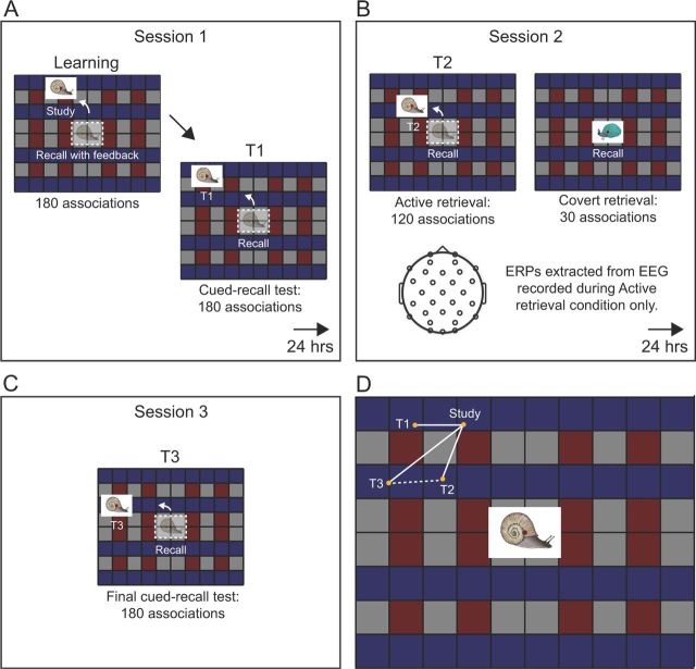
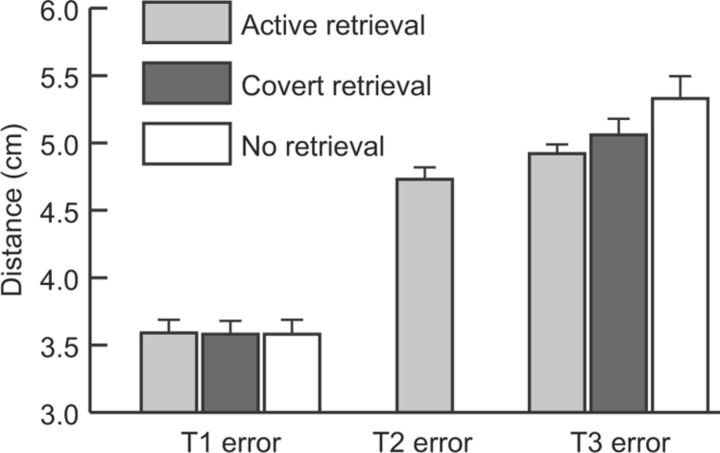
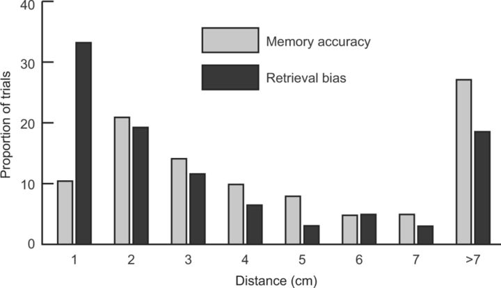
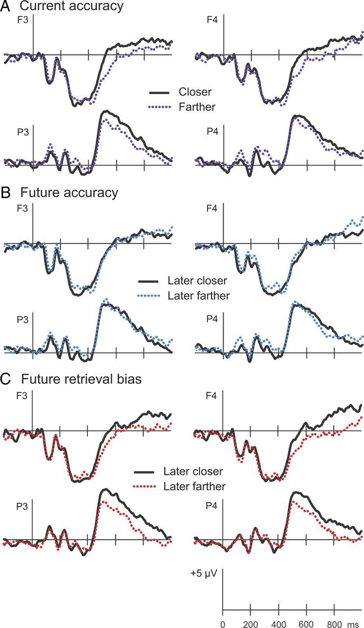
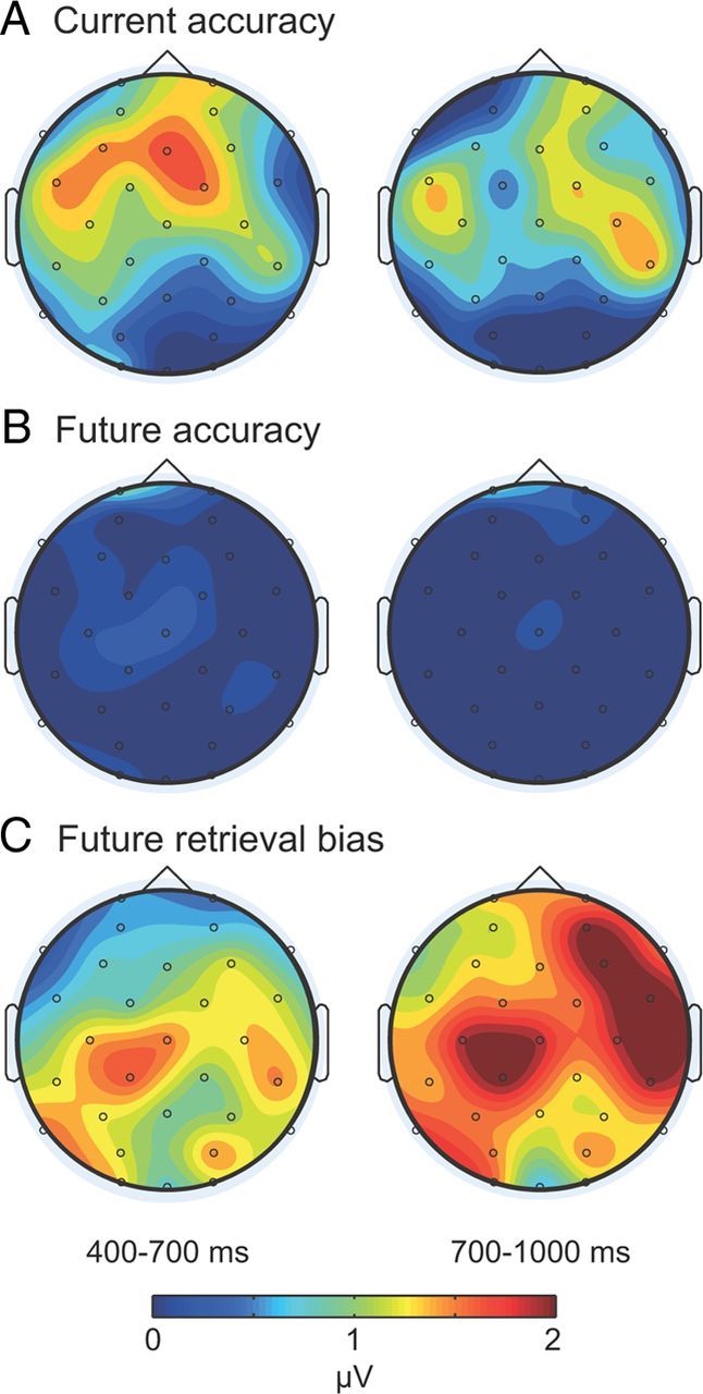
Reactivation of recently acquired information can strengthen memory storage and likely contributes to memory consolidation. Retrieval (generating information about prior events) may improve memory storage because it entails reactivation. Alternatively, retrieval may promote storage of retrieved information, and, if retrieval is inaccurate, subsequent recall could be distorted by the retrieved information. If retrieval modifies memory storage, as hypothesized, neural signals associated with accurate retrieval at that time may be distinct from neural signals associated with the degree of repeated retrieval error evident at some later time. We tested this prediction using a 3-session protocol. During session 1, people learned object-location associations to criterion and completed a cued-recall test in which locations were recalled upon viewing objects. During session 2, an electroencephalogram (EEG) was recorded during cued recall for a subset of the associations. During session 3, cued recall was tested for all associations. Retrieval improved storage, in that recall at session 3 was superior for objects tested in session 2 compared with those not tested. Retrieval-induced distortion was revealed in session 3 for those objects tested in session 2, in that those objects were generally placed closer to locations retrieved at session 2 relative to original study locations. EEG analyses revealed positive potentials (400-700 ms) associated with relatively accurate recall at session 2. Memory updating was reflected in positive potentials after 700 ms that differentially predicted the degree to which recall promoted storage of the session-2-retrieved location. These findings demonstrate unique neurocognitive processing whereby memories are updated with information produced during retrieval.


Figures

Figure 1.
Figure 1.
Schematic of the spatial-association task. A, At the beginning of the session-1 learning phase, each object was initially presented in a unique screen location on a 1024 × 768 pixel (26.67 × 20 cm) grid, viewed from a distance of 92 cm. After all learning trials, participants completed the first cued-recall test (T1). In this test, objects were shown in the center of the screen and participants were prompted to move each object to its original location. B, At session 2, EEG was recorded while participants completed the second cued-recall test (T2). For the Active condition, participants viewed each object for 1500 ms and then were prompted to move the object to its original location. For the Covert condition, participants prepared to recall the original location as in the Active condition, and likely attempted to retrieve the location during this time, but they had no opportunity to move the object. Trials in the Active and Covert conditions were randomly intermixed. C, At session 3, participants completed the final cued-recall test (T3). D, The original location for one example object in the Active condition is labeled “Study.” Solid white lines depict the distance the object was placed from the study location at each test. Typical errors made on each of the three tests as shown: mean error of 3.59 cm at T1, mean error of 4.73 cm at T2 (Active condition only), and mean error of 5.01 cm at T3. The dashed white line depicts the distance the object was placed from the T2 retrieved location at the final test (retrieval bias distance).
Figure 2.
Figure 2.
Mean errors in the spatial-association task. Errors on each test (T1, T2, and T3) were computed as the distance objects were placed from corresponding study locations. At T3, forgetting across days was significantly less for objects in the Active Retrieval condition versus the No Retrieval condition. Over all three conditions, errors were greater on T3 than on T1 (t(11) = 8.77, p < 0.001). For the Active Retrieval condition, errors were greater on T2 than on T1 (t(11) = 5.07, p < 0.001) and greater on T3 than on T2 (t(11) = 2.42, p < 0.05). Error bars show SEM after removing across-subject variability.
Figure 3.
Figure 3.
Retrieval bias contrasted with memory accuracy for the Active condition. Locations recalled at T3 were 4.92 cm (SE = 0.06) from the study location on average (memory accuracy) and 3.59 cm (SE = 0.06) from the T2 retrieved location on average (retrieval bias). Bins correspond to the proportion of objects placed fewer than the indicated number of centimeters from the study location or from the T2 retrieved location. Recalled locations were within 1 cm of the T2 retrieved location much more often then they were within 1 cm of the study location.
Figure 4.
Figure 4.
Electrophysiological results. ERPs recorded at session 2 were computed for objects in the Active condition placed closer to (solid lines) or farther from (dotted lines) the corresponding study or T2 locations. A, Current accuracy reflects distance objects were placed from the original location at T2. B, Future accuracy reflects distance objects were placed from the original location at T3. C, Future retrieval bias reflects distance objects were placed from the T2 retrieved location at T3. ERPs from two frontal (F3 and F4) and two parietal (P3 and P4) locations are displayed.
Figure 5.
Figure 5.
Topographic maps. A–C, Individual maps depict mean amplitude differences at 400–700 ms and 700–1000 ms for the contrast current accuracy (A), the contrast future accuracy (B), and the contrast future retrieval bias (C). Whereas differences tended to be relatively larger at frontal locations for the current accuracy contrast at 400–700 ms, differences were pronounced at most scalp locations at 700–1000 ms for the future retrieval bias contrast.

References

    1. Alvarez P, Squire LR. Memory consolidation and the medial temporal lobe: a simple network model. Proc Natl Acad Sci U S A. 1994;91:7041–7045. - PMC - PubMed
    1. Buckner RL, Wheeler ME, Sheridan MA. Encoding processes during retrieval tasks. J Cogn Neurosci. 2001;13:406–415. - PubMed
    1. Bunsey M, Eichenbaum H. Conservation of hippocampal memory function in rats and humans. Nature. 1996;379:255–257. - PubMed
    1. Butler AC, Marsh EJ, Goode MK, Roediger HL., III When additional multiple-choice lures aid versus hinder later memory. Appl Cogn Psychol. 2006;20:941–956.
    1. Carr MF, Jadhav SP, Frank LM. Hippocampal replay in the awake state: a potential substrate for memory consolidation and retrieval. Nat Neurosci. 2011;14:147–153. - PMC - PubMed

Publication types

LinkOut - more resources