Review
. 2012:2012:949157.
doi: 10.1155/2012/949157. Epub 2012 Aug 9.

Innate immune cells in liver inflammation

Affiliations
Review

Innate immune cells in liver inflammation

Evaggelia Liaskou et al. Mediators Inflamm. 2012.

Abstract

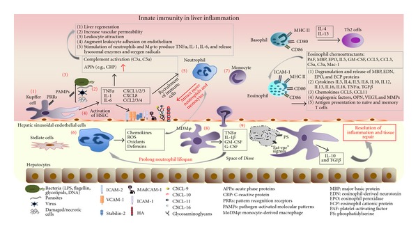
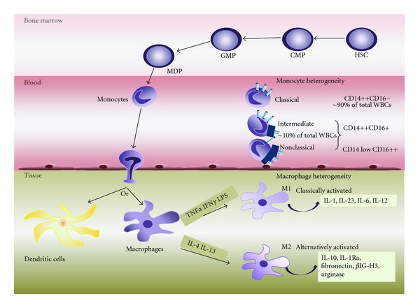
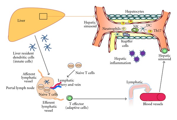
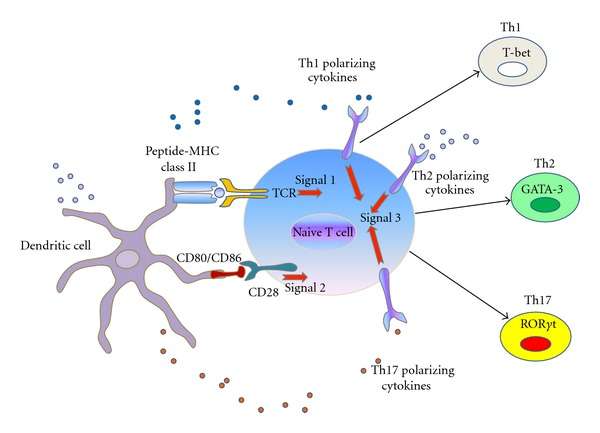
Innate immune system is the first line of defence against invading pathogens that is critical for the overall survival of the host. Human liver is characterised by a dual blood supply, with 80% of blood entering through the portal vein carrying nutrients and bacterial endotoxin from the gastrointestinal tract. The liver is thus constantly exposed to antigenic loads. Therefore, pathogenic microorganism must be efficiently eliminated whilst harmless antigens derived from the gastrointestinal tract need to be tolerized in the liver. In order to achieve this, the liver innate immune system is equipped with multiple cellular components; monocytes, macrophages, granulocytes, natural killer cells, and dendritic cells which coordinate to exert tolerogenic environment at the same time detect, respond, and eliminate invading pathogens, infected or transformed self to mount immunity. This paper will discuss the innate immune cells that take part in human liver inflammation, and their roles in both resolution of inflammation and tissue repair.


Figures

Figure 1
Figure 1
Innate immune cells in liver inflammation. During an infectious insult in the liver (1) resident macrophages, Kupffer cells, are the first immune cells to detect the presence of invading pathogens (bacteria, parasites, viruses, damaged, and/or necrotic cells) via PRRs/PAMPs. (2) Upon activation Kupffer cells release cytokines TNFα, IL-1, and IL-6 as well as chemokines CXCL 1–3, CXCL-8, CCL-2–4 that initiate (3) the acute-phase response and inflammation. Acute inflammation is characterized by the rise in plasma proteins, collectively named acute-phase proteins (APPs) that include C-reactive protein (CRP) and complement components. (4) Proinflammatory cytokines released from activated Kupffer cells can activate hepatic sinusoidal endothelial cells to upregulate adhesion molecules (ICAM1 and 2, VCAM-1, MAdCAM etc.) and in combination with the chemokines secreted from Kupffer cells can stimulate the recruitment of neutrophils and monocytes to the liver. (5) Neutrophils are the initial phagocytes to arrive at the site of microbial invasion, where (6) they change their phenotype, they become activated and release powerful and cytotoxic antimicrobial molecules such as reactive oxygen species (ROS), oxidants, defensins, as well as chemokines to attract more neutrophils and monocytes. (7) Following their recruitment to the tissue, monocytes undergo differentiation into (8) tissue macrophages (MDMφ), which release TNFα, IL-1β, G-CSF, and GM-CSF factors that can extend the lifespan of neutrophils thus sustaining their presence at the site of inflammation. (9) In order for inflammation to be resolved, the dangerous neutrophils at the inflammatory loci undergo apoptosis and terminate the inflammatory process quickly. Apoptotic neutrophils represent an important anti-inflammatory stimulus to other cells involved in the resolution of inflammation by producing “eat-me” signals recognised by the surrounding phagocytes. Phosphatidylserine (PS) residues on the apoptotic neutrophil membrane allow recognition by its receptor on macrophages, which not only initiates phagocytosis but also modifies the transcriptional profile of the Mφ, increasing the production of IL-10 and TGF-b, cytokines associated with resolution of inflammatory response and tissue repair. Basophils are short-lived cells that express MHC II and CD80/CD86 costimulatory molecules, thus are able to present antigens to CD4+ T cells promoting their differentiation into Th2 cells via release of IL-4 and IL-13. Eosinophils recruited to the liver release proinflammatory mediators including granule-stored cationic proteins, cytokines, and chemokines. They also express MHC II, CD80/CD86, CD40, and ICAM-1; thus they are able to present antigens to T cells initiating or amplifying antigenic-specific immune responses.
Figure 2
Figure 2
Monocyte and macrophage heterogeneity. Monocytes originate in the bone marrow where they develop from hematopoietic stem cells (HSCs) via several differentiation steps and intermediate progenitor stages that pass through the common myeloid progenitor (CMP), the granulocyte/macrophage progenitor (GMP), and the macrophage/DC progenitor (MDP) stages. The MDP gives rise to monocytes, which are released in blood circulation where they remain for 1–3 days. In peripheral blood, circulating monocytes represent ~5–10% of peripheral blood white blood cells (WBCs) and are a highly heterogenic population. Three main subtypes have been described based on the expression of CD14 and CD16 receptors: the classical CD14++CD16, intermediate CD14++CD16+, and nonclassical CD14 low CD16++ monocytes. In general, circulation monocytes are recruited to tissues where they can differentiate into dendritic cells or tissue macrophages (Kupffer cells in the liver; microglial cells in the brain, etc.), replenishing the existing populations. Additional heterogeneity also exists between the macrophages, with two major classes being identified: the classically activated (M1) and the alternatively activated (M2) macrophages. M1 macrophages are developed in response to TNFα and IFNγ as well as in response to microbial products such as LPS, and they produce in turn proinflammatory cytokines including IL-1, IL-23, IL-6, and IL-12. M2 macrophages can develop in response to IL-4 and IL-13 cytokines and play important roles in down-regulation of inflammation and tissue remodelling by releasing IL-10 and IL-1 receptor antagonist (IL-1Ra). They also produce high levels of arginase, fibronectin, and a matrix-associated protein, βIG-H3.
Figure 3
Figure 3
Innate immune cell (neutrophils, NK cells and monocytes) recruitment to hepatic inflammation. Human liver receives dual blood supply from both portal vein and hepatic artery. During the inital event of hepatic inflammation, innate immune cells such as neutrophils, monocytes and NK cells are recruited to the liver. Liver resident dendritic cells sample the foreign antigen and carry to local draining portal lymph nodes where antigens are presented to the adaptive naive T cells. Following the antigen presentation, different types of antigen-specific T effectors cells leave the nodes and drain back to systemic circulation. These T effector cells recruit via hepatic sinusoid towards the site of injury or inflammation. Th17 cells which secrete IL-17 attract neutrophils and also link innate and adaptive immunity.
Figure 4
Figure 4
Linking innate and adaptive immune system. Dendritic cells from innate immune system present their antigen to naive T cells at local draining lymph nodes. T-cell receptor (TCR) ligation to MHC class II associated peptides processed from pathogens (signal 1) and binding of costimulatory molecule CD28 on lymphocyte to CD80 and CD86 expressed by dendritic cells (signal 2) leads to T-cell lineages differentiation. Signal 3 is the polarizing cytokines signals from the innate immune cells. Depending on type of antigen which is presented and nature of cytokines in the microenvironment, innate DC cells direct the development of Th1, Th2, Th17 lymphocytes lineages which plays crucial role in adaptive immune system.

References

    1. O’Farrelly C, Doherty DG. Liver Immunology Principles and Practice. chapter 1. 2007. A short primer on fundamental immunology; pp. 15–24.
    1. Triantafilou M, Lepper PM, Olden R, Dias IS, Triantafilou K. Location, location, location: is membrane partitioning everything when it comes to innate immune activation? Mediators of Inflammation. 2011;2011:10 pages.186093 - PMC - PubMed
    1. Akira S, Uematsu S, Takeuchi O. Pathogen recognition and innate immunity. Cell. 2006;124(4):783–801. - PubMed
    1. Lalor PF, Adams DH. The liver: a model of organ-specific lymphocyte recruitment. Expert Reviews in Molecular Medicine. 2002;4(2):1–16. - PubMed
    1. Gao B, Jeong WI, Tian Z. Liver: an organ with predominant innate immunity. Hepatology. 2008;47(2):729–736. - PubMed

Publication types

LinkOut - more resources