. 2012:18:2221-8.
Epub 2012 Aug 9.

Cytopathic effect of Acanthamoeba on human corneal fibroblasts

Affiliations

Cytopathic effect of Acanthamoeba on human corneal fibroblasts

Noriko Takaoka-Sugihara et al. Mol Vis. 2012.

Abstract

Purpose: Acanthamoeba keratitis is associated with keratocyte depletion in humans. We investigated how Acanthamoebae isolated from corneas affected by Acanthamoeba keratitis interacted with human corneal stromal cells in vitro.

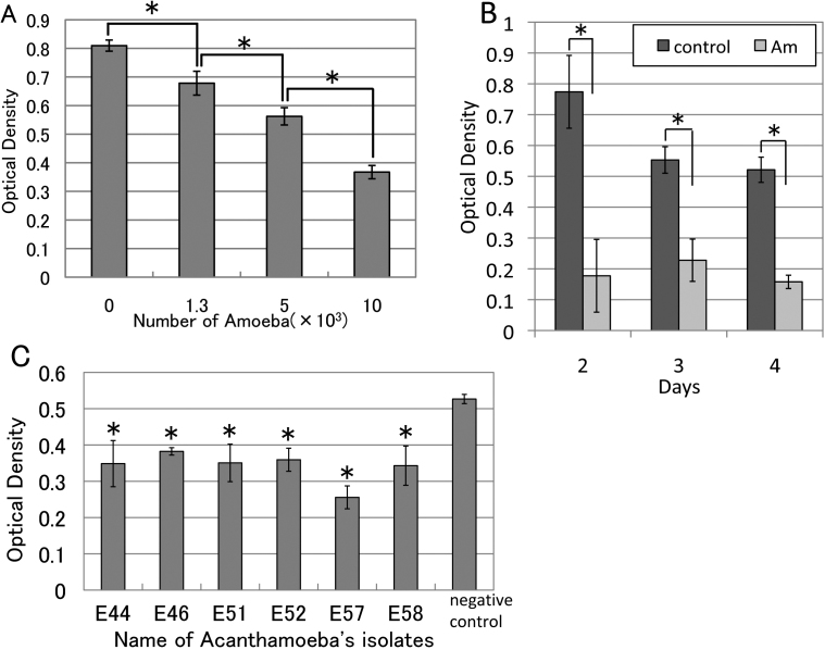
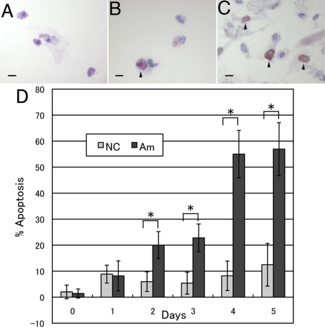
Methods: Acanthamoebae were isolated from 6 patients with Acanthamoeba keratitis and genotyping was done. Whether the isolated Acanthamoebae could invade the corneal stroma was assessed with denuded corneal stroma ex vivo. The cytopathic effect of Acanthamoeba on cultured corneal fibroblasts from donor corneas was quantitatively evaluated by the MTT assay after culture under various conditions. Terminal deoxynucleotidyl transferase-mediated dUTP nick-end labeling (TUNEL) and Annexin V staining were employed to detect apoptotic cells among the corneal fibroblasts co-cultured with Acanthamoebae.

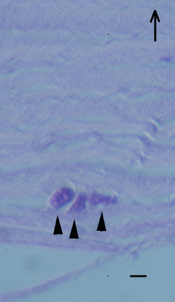
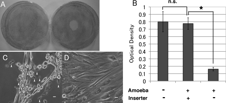
Results: All 6 Acanthamoebae isolated from the patients with Acanthamoeba keratitis were shown to have the T4 genotype by 18S rDNA sequence analysis. Acanthamoebae invaded the denuded corneal stroma in the ex vivo experiments and had a cytopathic effect on human corneal fibroblasts after direct adhesion, but not via chemical mediators. A cytopathic effect was detected with all 6 Acanthamoebae and corneal fibroblasts mainly died by apoptosis, as evidenced by Annexin V staining.

Conclusions: Acanthamoebae isolated from patients with Acanthamoeba keratitis had a cytopathic effect on human corneal fibroblasts, mainly via induction of apoptosis after direct adhesion. Our findings may provide some clues to the pathophysiology of corneal keratocyte depletion in patients with Acanthamoeba keratitis.


Figures

Figure 1
Figure 1
Ex vivo invasion of Acanthamoeba into corneal stroma. Acanthamoebae were added to denuded human corneal stroma. Acanthamoebae were placed on the denuded corneal stroma (endothelial side up) and incubated at 25 °C for 2 days. Hematoxylin and eosin staining shows Acanthamoebae (arrowheads) located in fine collagen fibrils. Arrow shows the direction of corneal epithelium. Scale bar=10 µm.
Figure 2
Figure 2
Direct and indirect cytopathic effects of Acanthamoeba on corneal fibroblasts. A: Acanthamoebae were placed on corneal fibroblasts at the center of 6-cm dishes and incubated at 25 °C for 2 days. The fibroblasts are uniformly stained with Giemsa solution in a control dish (left). The central area where Acanthamoebae were placed shows no staining (indicating loss of corneal fibroblasts) in a treated dish (right). B: MTT assay showed there was no significant difference of optical density value in the outer dishes with corneal fibroblasts with or without insert culture dishes bearing Acanthamoebae. Significant low optical density value is detected in Acanthamoebae direct adhesion group, compared with insert culture dishes bearing Acanthamoebae. (n=6) Amoeba; Acanthamoeba, Inserter; insert culture dish. C: Phase contrast microscopy shows many corneal fibroblasts are detached and Acanthamoebae adhere to corneal fibroblasts and the dish surface. Arrowheads show active Acanthamoebae co-cultured with corneal fibroblasts. D: Confluent human corneal fibroblasts are seen. Acanthamoebae in the insert culture dishes with 0.4 µm pores are not observed. Similar findings were obtained with repeated two sets of experiments. Representative data are shown. Scale bar=10 µm.
Figure 3
Figure 3
Cytopathic effect of Acanthamoeba on corneal fibroblasts in various conditions. A: Acanthamoebae (0 to 10×103) were added to corneal fibroblasts in each well and incubated at 25 °C for 2 days. Acanthamoebae (1×10⁴) significantly decreased the viability of corneal fibroblasts compared with no Acanthamoebae (n=4). B: A significant decrease of corneal fibroblast viability was detected from day 2 (n=4). C: Cytopathic effect on corneal fibroblasts for 6 Acanthamoebae isolates from our AK patients. A significant decrease of optical density (indicating a cytopathic effect on corneal fibroblasts) was detected with all tested Acanthamoebae compared to control cultures with no Acanthamoebae (n=4). Similar findings were obtained with repeated two experiments. Representative data are shown. *p<0.05.
Figure 4
Figure 4
Detection of apoptotic human corneal fibroblasts. TUNEL staining was performed after human corneal fibroblasts were co-cultured with Acanthamoebae. TUNEL-positive cells are not detected in cultures of fibroblasts without Acanthamoebae (A). Arrowheads show TUNEL-positive apoptotic corneal fibroblasts cultured with Acanthamoebae (B) and with Actinomysin D (C). Scale bar=100 µm. D: Percentage of Annexin V-positive cells. A significant increase of Annexin V-positive corneal fibroblasts was detected on day 2 or later of culture with Acanthamoebae. More than 50% of corneal fibroblasts were Annexin V-positive on days 4 and 5 (n=4–5). Similar results were obtained with repeated two experiments. NC; Negative control, corneal fibroblasts without Acanthamoebae, Am; Corneal fibroblasts co-cultured with Acanthamoebae. *p<0.05.

References

    1. Illingworth CD, Cook SD. Acanthamoeba keratitis. Surv Ophthalmol. 1998;42:493–508. - PubMed
    1. Dart JK, Saw VP, Kilvington S. Acanthamoeba keratitis: diagnosis and treatment update 2009. Am J Ophthalmol. 2009;148:487–99. - PubMed
    1. Stehr-Green JK, Bailey TM, Visvesvara GS. The epidemiology of Acanthamoeba keratitis in the United States. Am J Ophthalmol. 1989;107:331–6. - PubMed
    1. Sharma S, Garg P, Rao GN. Patient characteristics, diagnosis, and treatment of non-contact lens related Acanthamoeba keratitis. Br J Ophthalmol. 2000;84:1103–8. - PMC - PubMed
    1. Dart JK, Saw VP, Kilvington S. Acanthamoeba keratitis: diagnosis and treatment update 2009. Am J Ophthalmol. 2009;148:487–99. - PubMed

Publication types

MeSH terms

LinkOut - more resources