

Infrahepatic caudal/inferior vena cava interruption with azygos/hemiazygos continuation. Vascular anomaly in swine
- PMID: 22933907
- PMCID: PMC3423700
- DOI: 10.2478/v10019-010-0029-5
Infrahepatic caudal/inferior vena cava interruption with azygos/hemiazygos continuation. Vascular anomaly in swine
Abstract
Background: Swine are commonly used as a model to study congenital cardiovascular defects that occur in humans and these models have been both spontaneous and experimentally induced. Ventricular septal defect, patent ductus arteriosus, and atrial septal defect (ASD) are examples of experimentally induced models. Absence of caudal/inferior vena cava (CVC/IVC) with azygos/hemiazygos continuation is an uncommon vascular anomaly.
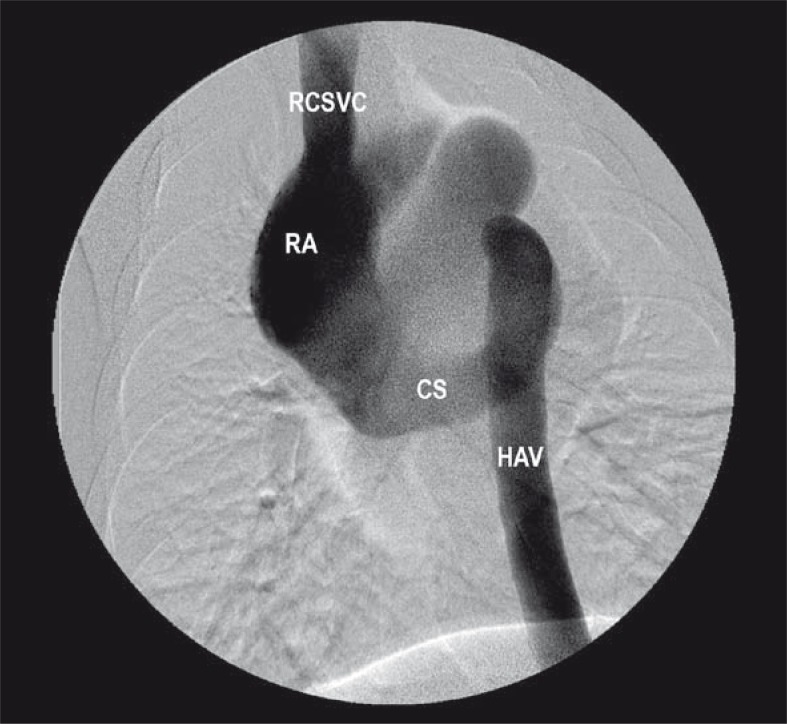
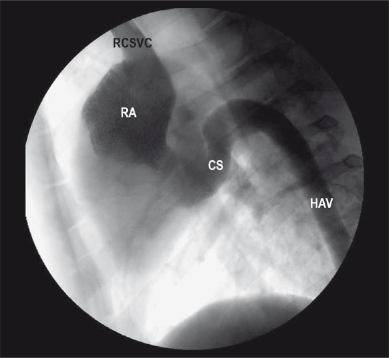
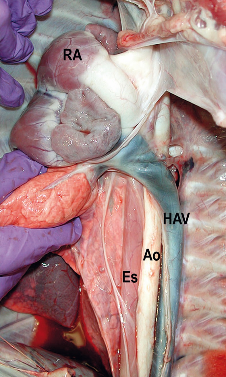
Case report: The vascular anomaly presented in this case report was an incidental finding on a pig that was evaluated for experimental percutaneous atrial septal defect creation and its closure using a percutaneous femoral vein approach. Absence of CVC/IVC was confirmed by venography and necropsy.
Conclusions: To the best of the investigators knowledge, this is the first report of absence of CVC/IVC with azygos/hemiazygos continuation in the swine.
Keywords: azygos vein; congenital vascular anomaly; experimental animal model; hemiazygos vein; inferior vena cava.
Figures
Similar articles
-
Percutaneous atrial septal defect closure through femoral and transjugular approaches in patients with interrupted inferior vena cava.J Cardiol Cases. 2018 Jun 9;18(3):106-109. doi: 10.1016/j.jccase.2018.05.007. eCollection 2018 Sep. J Cardiol Cases. 2018. PMID: 30279924 Free PMC article.
-
Totally endoscopic atrial septal defect repair in a patient with interrupted inferior vena cava with azygos-hemiazygos continuation.J Card Surg. 2021 Dec;36(12):4814-4817. doi: 10.1111/jocs.16036. Epub 2021 Sep 27. J Card Surg. 2021. PMID: 34570371
-
Double Inferior Vena Cava (IVC) With Intrahepatic Interruption, Hemiazygos Vein Continuation, and Intrahepatic Venous Shunt.Vasc Endovascular Surg. 2017 Jan;51(1):38-42. doi: 10.1177/1538574416687734. Vasc Endovascular Surg. 2017. PMID: 28100158
-
Liver transplantation in a patient with developmental interruption of the inferior vena cava with azygos substitution.Transplant Proc. 2012 Jun;44(5):1460-3. doi: 10.1016/j.transproceed.2012.01.121. Transplant Proc. 2012. PMID: 22664037 Review.
-
[Congenital anomaly of the inferior vena cava with hemiazygos continuation. Ultrasonic diagnosis].Ann Radiol (Paris). 1990;33(6):339-46. Ann Radiol (Paris). 1990. PMID: 2085271 Review. French.
Cited by
-
Double BioDisk: a new bioprosthetic device for transcatheter closure of atrial septal defects - a feasibility study in adult sheep.Radiol Oncol. 2012 Jun;46(2):89-96. doi: 10.2478/v10019-012-0029-8. Epub 2012 May 30. Radiol Oncol. 2012. PMID: 23077444 Free PMC article.
-
Slovenian experience from diagnostic angiography to interventional radiology.Radiol Oncol. 2014 Nov 5;48(4):416-25. doi: 10.2478/raon-2014-0023. eCollection 2014 Dec. Radiol Oncol. 2014. PMID: 25435857 Free PMC article.
-
Iodine based radiopacity of experimental blood clots for testing of mechanical thrombectomy devices.Radiol Oncol. 2013 Mar;47(1):14-8. doi: 10.2478/raon-2013-0001. Epub 2013 Feb 1. Radiol Oncol. 2013. PMID: 23450088 Free PMC article.
References
-
- Swindle MM, Thompson RP, Carabello BA, Smith AC, Green CT, Gillette PC. Congenital cardiovascular disease. In: Swindle MM, editor. Swine as models in biomedical research. Ames: Iowa State University Press, Inc; 1992. pp. 176–84.
-
- Wei Lu, Pavcnik D, Uchida B, Park WK, Liu L, Timmermans HA, et al. The ovine jugular vein as a model for interventional radiology procedures. Radiol Oncol. 2008;42:59–65.
-
- Dondelinger RF, Ghysels MP, Brisbois D, Donkers E, Snaps FR, Saunders J, et al. Relevant radiological anatomy of the pig as trining model in interventional radiology. Eur Radiol. 1998;8:1254–73. - PubMed
-
- Miller ME. Veins. In: Evans HE, Christensen GC, editors. Miller’s anatomy of the dog. 2th edition. Philadelphia: Saunders; 1979. pp. 757–801.
-
- Kutlu R. Obliterative hepatocavopathy – ultrasound and cavography findings. Radiol Oncol. 2008;42:181–6.
LinkOut - more resources
Full Text Sources