

Radiological considerations in von Hippel-Lindeau disease: imaging findings and the review of the literature
- PMID: 22933910
- PMCID: PMC3423690
- DOI: 10.2478/v10019-010-0014-z
Radiological considerations in von Hippel-Lindeau disease: imaging findings and the review of the literature
Abstract
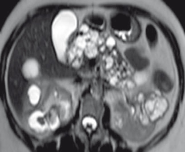
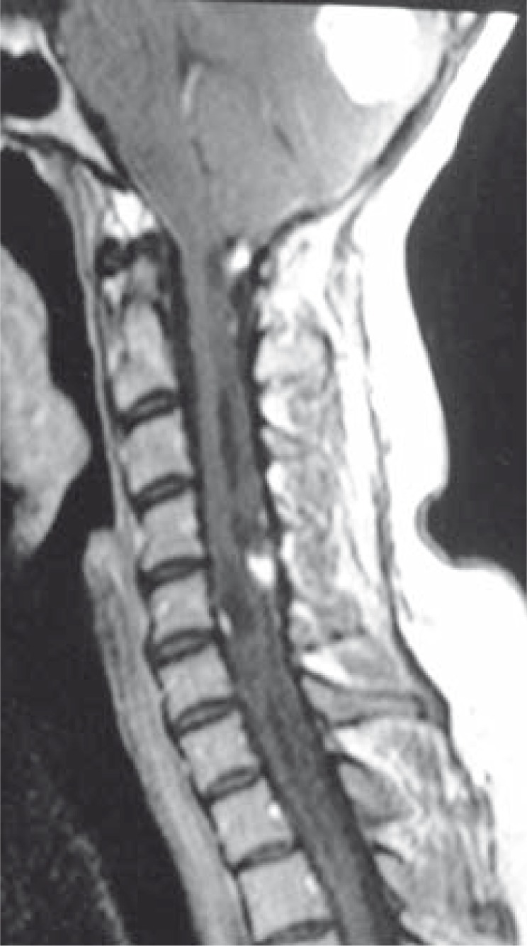
Background: Von Hippel Lindau disease is an autosomal dominant multisystem/multitumoral cancer disease diagnosed by clinical, radiologic and genetic findings. Its prevalence has been estimated to be of 1/36000 inhabitants. The tumours can be benign or malignant.
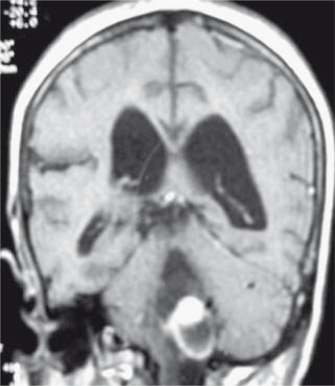
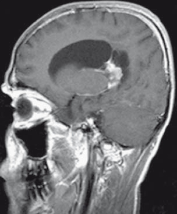
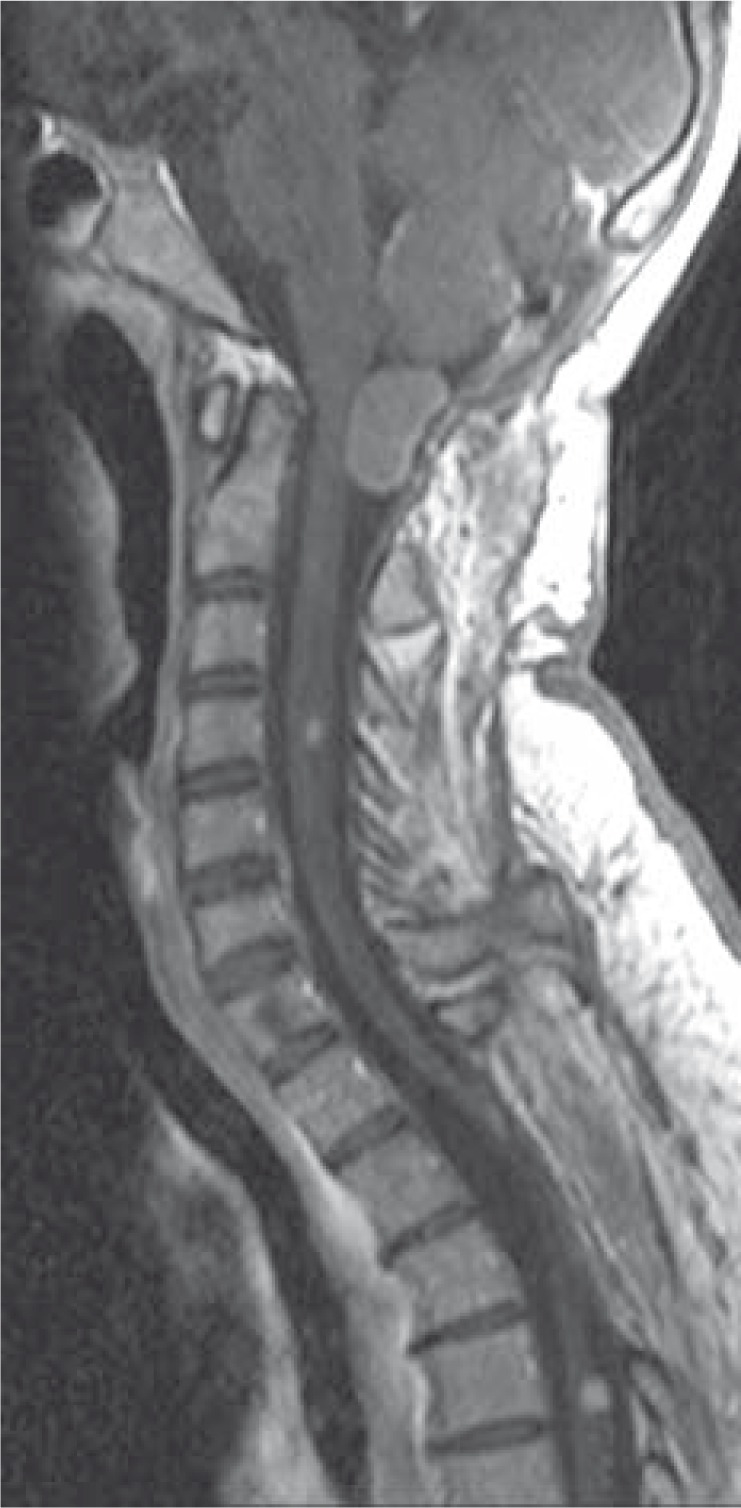
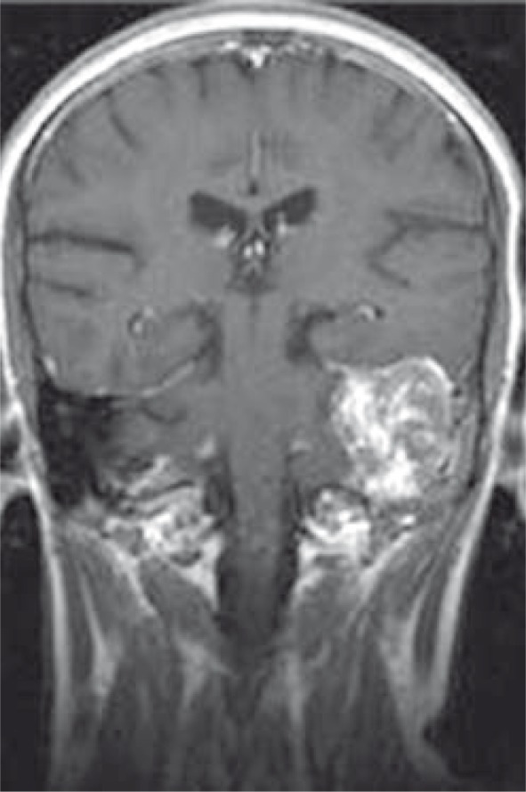
Case report: We represent MR findings of a family with ten children. Mother and five siblings had von Hippel-Lindau disease.
Conclusions: Radiologic imaging is very important for the early diagnosis and treatment of asymptomatic patients. Diagnosing it early is important because the tumours in von Hippel Lindau disease are treatable. Also, an early detection allows the patient's survival and quality of life. A multidisciplinary team approach is important in screening.
Keywords: brain; magnetic resonance imaging; spine; tumours; von Hippel-Lindau.
Figures
Similar articles
-
Imaging features of von Hippel-Lindau disease.Radiographics. 2008 Jan-Feb;28(1):65-79; quiz 323. doi: 10.1148/rg.281075052. Radiographics. 2008. PMID: 18203931 Review.
-
Bilateral pheochromocytoma as first manifestation of von Hippel-Lindau disease: a case report.Turk J Pediatr. 2012 Sep-Oct;54(5):532-5. Turk J Pediatr. 2012. PMID: 23427520
-
The impact of molecular genetic analysis of the VHL gene in patients with haemangioblastomas of the central nervous system.J Neurol Neurosurg Psychiatry. 1999 Dec;67(6):758-62. doi: 10.1136/jnnp.67.6.758. J Neurol Neurosurg Psychiatry. 1999. PMID: 10567493 Free PMC article.
-
Von Hippel - Lindau disease.Malays Fam Physician. 2017 Apr 30;12(1):29-31. eCollection 2017. Malays Fam Physician. 2017. PMID: 28503272 Free PMC article.
-
VON HIPPEL-LINDAU DISEASE: Update on Pathogenesis and Systemic Aspects.Retina. 2019 Dec;39(12):2243-2253. doi: 10.1097/IAE.0000000000002555. Retina. 2019. PMID: 31095066 Review.
Cited by
-
Multidisciplinary management of patients diagnosed with von Hippel-Lindau disease: A practical review of the literature for clinicians.Asian J Urol. 2022 Oct;9(4):430-442. doi: 10.1016/j.ajur.2022.08.002. Epub 2022 Sep 10. Asian J Urol. 2022. PMID: 36381595 Free PMC article. Review.
-
Assessing renal function in children with hydronephrosis - additional feature of MR urography.Radiol Oncol. 2011 Dec;45(4):248-58. doi: 10.2478/v10019-011-0038-z. Epub 2011 Nov 16. Radiol Oncol. 2011. PMID: 22933962 Free PMC article.
References
-
- Shuin T, Yamasaki I, Tamura K, Okuda H, Furihata M, Ashida S. Von Hippel-Lindau disease: molecular pathological basis, clinical criteria, genetic testing, clinical features of tumors and treatment. Jpn J Clin Oncol. 2006;6:337–43. - PubMed
-
- Latif F, Tory K, Gnarra J, Yao M, Duh FM, Orcutt ML, et al. Identification of the von Hippel-Lindau disease tumor suppressor gene. Science. 1993;260:1317–20. - PubMed
-
- Melmon KL, Rosen SW. Lindau’s disease: review of the literature and study of a large kindred. Am J Med. 1964;36:595–617. - PubMed
-
- Leung RS, Biswas SV, Duncan M, Rankin S. Imaging features of von Hippel-Lindau disease. Radiographics. 2008;28:65–79. - PubMed
-
- Meister M, Choyke P, Anderson C, Patel U. Radiological evaluation, management, and surveillance of renal masses in Von Hippel-Lindau disease. Clin Radiol. 2009;6:589–600. - PubMed
LinkOut - more resources
Full Text Sources