. 2011 Dec;45(4):259-66.
doi: 10.2478/v10019-011-0034-3. Epub 2011 Oct 8.

Cathepsin H indirectly regulates morphogenetic protein-4 (BMP-4) in various human cell lines

Affiliations

Cathepsin H indirectly regulates morphogenetic protein-4 (BMP-4) in various human cell lines

Matija Rojnik et al. Radiol Oncol. 2011 Dec.

Abstract

Background: Cathepsin H is a cysteine protease considered to play a major role in tumor progression, however, its precise function in tumorigenesis is unclear. Cathepsin H was recently proposed to be involved in processing of bone morphogenetic protein 4 (BMP-4) in mice. In order to clarify whether cathepsin H also regulates BMP-4 in humans, its impact on BMP-4 expression, processing and degradation was investigated in prostate cancer (PC-3), osteosarcoma (HOS) and pro-monocytic (U937) human cell lines.

Materials and methods: BMP-4 expression was founded to be regulated by cathepsin H using PCR array technology and confirmed by real time PCR. Immunoassays including Western blot and confocal microscopy were used to evaluate the influence of cathepsin H on BMP-4 processing.

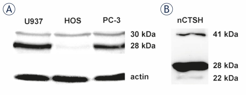
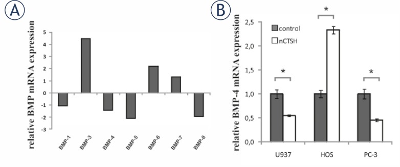
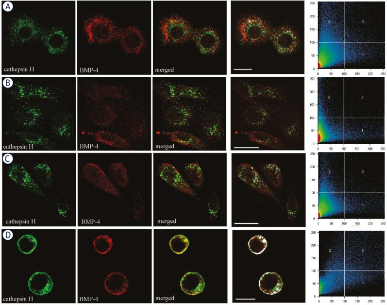
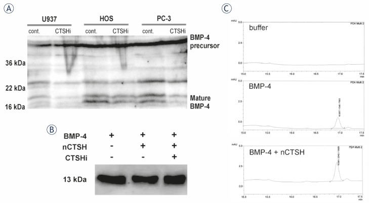
Results: In contrast to HOS, the expression of BMP-4 mRNA in U937 and PC3 cells was significantly decreased by cathepsin H. The different regulation of BMP-4 synthesis could be associated with the absence of the mature 28 kDa cathepsin H form in HOS cells, where only the intermediate 30 kDa form was observed. No co-localization of BMP-4 and cathepsin H was observed in human cell lines and the multistep processing of BMP-4 was not altered in the presence of specific cathepsin H inhibitor. Isolated cathepsin H does not cleave mature recombinant BMP-4, neither with its amino- nor its endopeptidase activity.

Conclusions: Our results exclude direct proteolytic processing of BMP-4 by cathepsin H, however, they provide support for its involvement in the regulation of BMP-4 expression.

Keywords: bone morphogenetic protein 4; cancer; cathepsin H; human cell lines; proteolytic enzymes.


Conflict of interest statement

Disclosure: No potential conflicts of interest were disclosed.

Figures

FIGURE 1
FIGURE 1
CTSH processing forms. (A) Different forms of CTSH were detected in human cell lines and (B) in the sample of nCTSH using Western blot. CTSH was visualized with sheep anti-CTSH primary pAb and rabbit anti-sheep horse radish peroxidise (HRP) labeled secondary antibody. The intermediate 30 kDa CTSH form is present in all of the selected cell lines, while the mature 28 kDa CTSH form is missing in HOS cells. A procathepsin H (41 kDa) and single chain form (22 kDa) could be detected in smaller amounts compared to the mature forms. nCTSH contains the procathepsin H, the mature and heavy chain forms.
FIGURE 2
FIGURE 2
CTSH dependent cytokine mRNA expression. (A) The effect of CTSH on the mRNA expression of cytokines from BMP family. Differentiated U937 cells were incubated with 0.5 μM nCTSH and the mRNA levels were screened by PCR-arrays enabling simultaneous screen of the of 84 cytokine genes. The expression of BMP-2 is not presented, while it was probably an artifact, as shown by the analysis of the melting curve. (B) The influence of CTSH on BMP-4 mRNA expression was further evaluated with quantitative real time PCR analysis in U937, HOS and PC-3 cell lines. Cells were treated with 0.5 μM nCTSH for 24 h. The mRNA levels obtained from control samples were normalized to 1. Each bar represents the mean±SD. Ns, non-significant, *P<0.05.
FIGURE 3
FIGURE 3
Co-localization of CTSH (green fluorescence) and BMP-4 (red fluorescence) in U937 (A), HOS (B), PC-3 (C) and WEHI231 (D) cells. Weak or no co-localization was found in selected human cells (A, B, C), while clear co-localization can be seen in mouse WEHI231 cell line (D). CTSH was labeled with 1D10 monoclonal primary antibody and anti-mouse AlexaFluor™488 secondary antibody. BMP-4 was labeled with primary goat polyclonal anti-BMP-4 antibody (Santa Cruz) and anti-goat AlexaFluor™ 555 secondary antibody. The sites of co-localization are shown in white (frame 4) and correspond to the pixels that are over the threshold in both channels (frame 5). Scale bars represent 5 μm (A and D) and 20 μm (B and C).
FIGURE 4
FIGURE 4
CTSH processing of mature BMP-4 protein. (A) Human cell lines were treated with 5 μM CTSHi for 24 h and then cell lysates were analyzed using Western blot. Proteins (50 μg) from cell lysates were separated on SDS-PAGE and transferred on to PVDF membrane. BMP-4 was detected with anti-BMP-4 N16 antibody (Santa Cruz) and then with secondary antibody labelled with HRP. The molecular mass in kDa is indicated on the left hand side of the blots. Molecular mass of the mature BMP-4 is detected to be approximately 18 kDa. (B) Using Western blot we analyzed the products of the reaction between mature human recombinant BMP-4 and nCTSH. Mature human recombinant BMP-4 was incubated for 1.5 h at 37ºC in CTSH activity buffer (lane 1), with 60 ng of nCTSH in CTSH activity buffer (lane 2) and 60 ng of nCTSH in CTSH activity buffer that was pre-treated for 10 min with 10 μM CTSHi (lane 3). Mature human recombinant BMP-4 has a molecular mass of 13 kDa. (C) Using reverse phase HPLC we analyzed CTSH activity buffer (buffer), mature human recombinant BMP-4 in CTSH activity buffer (BMP-4) and the products of the reaction between mature human recombinant BMP-4 and nCTSH in CTSH activity buffer (BMP-4 + nCTSH). BMP-4 was eluted in the fraction around 17.0 min.

Similar articles

Cited by

References

    1. Turk V, Turk B, Turk D. Lysosomal cysteine proteases: facts and opportunities. EMBO J. 2001;20:4629–33. - PMC - PubMed
    1. Kirschke H, Cathepsin H. In: Handbook of proteolytic enzymes. 2nd edition. Barret AJ, Rawlings ND, Woessner JF, editors. London: Elsevier Academic Press; 2004. pp. 1089–92.
    1. Kirschke H, Barrett AJ, Rawlings ND. Lysosomal cysteine proteases. 2nd edition. Oxford: Oxford University Press; 1998.
    1. Ritonja A, Popovic T, Kotnik M, Machleidt W, Turk V. Amino acid sequences of the human kidney cathepsins H and L. FEBS Lett. 1988;228:341–5. - PubMed
    1. Vasiljeva O, Dolinar M, Turk V, Turk B. Recombinant human cathepsin H lacking the mini chain is an endopeptidase. Biochemistry. 2003;42:13522–8. - PubMed

LinkOut - more resources