

LRP-1 and LRP-2 receptors function in the membrane neuron. Trafficking mechanisms and proteolytic processing in Alzheimer's disease
- PMID: 22934024
- PMCID: PMC3429044
- DOI: 10.3389/fphys.2012.00269
LRP-1 and LRP-2 receptors function in the membrane neuron. Trafficking mechanisms and proteolytic processing in Alzheimer's disease
Abstract
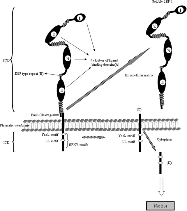
Low density lipoprotein receptor-related protein (LRP) belongs to the low-density lipoprotein receptor family, generally recognized as cell surface endocytic receptors, which bind and internalize extracellular ligands for degradation in lysosomes. Neurons require cholesterol to function and keep the membrane rafts stable. Cholesterol uptake into the neuron is carried out by ApoE via LRPs receptors on the cell surface. In neurons the most important are LRP-1 and LRP-2, even it is thought that a causal factor in Alzheimer's disease (AD) is the malfunction of this process which cause impairment intracellular signaling as well as storage and/or release of nutrients and toxic compounds. Both receptors are multifunctional cell surface receptors that are widely expressed in several tissues including neurons and astrocytes. LRPs are constituted by an intracellular (ICD) and extracellular domain (ECD). Through its ECD, LRPs bind at least 40 different ligands ranging from lipoprotein and protease inhibitor complex to growth factors and extracellular matrix proteins. These receptors has also been shown to interact with scaffolding and signaling proteins via its ICD in a phosphorylation-dependent manner and to function as a co-receptor partnering with other cell surface or integral membrane proteins. Thus, LRPs are implicated in two major physiological processes: endocytosis and regulation of signaling pathways, which are both involved in diverse biological roles including lipid metabolism, cell growth processes, degradation of proteases, and tissue invasion. Interestingly, LRPs were also localized in neurons in different stages, suggesting that both receptors could be implicated in signal transduction during embryonic development, neuronal outgrowth or in the pathogenesis of AD.
Keywords: Alzheimer's disease; LRP-1; LRP-2; amyloid-beta; astrocytes; brain; central nervous system; intracellular domain; megalin; neurodegenerative diseases; neuron.
Figures

References
-
- Ambjorn M., Asmussen J. W., Lindstam M., Gotfryd K., Jacobsen C., Kiselyov V. V., Moestrup S., Penkova M., Bock E., Berezin V. (2008). Metallothionein and a peptide modelled after metallothionein, Emtin, B, induced neuronal differentiation and survival through binding to receptors of the low-density lipoprotein receptor family. J. Neurochem. 104, 21–37 10.1111/j.1471-4159.2007.05036.x - DOI - PubMed
-
- Ananyeva N. M., Makogonenko Y. M., Sarafanov A. G., Pechik I. V., Gorlatova N., Radtke K. P., Shima M., Saenko E. L. (2008). Inteaction of coagulation factor VIII with members of the low-density lipoprotein receptor family follows common mechanism and involves consensus residues within the A2 binding site 484–509. Blood Coagul. Fibrinolysis 19, 543–555 10.1097/MBC.0b013e3283068859 - DOI - PubMed
-
- Assemat E., Chatelet F., Chandellier J., Commo F., Cases O., Verroust P., Kozyraki R. (2005). Overlapping expression patterns of the multiligand endocyte receptors cubilin and megalin in the CNS, sensory organs and developing epithelia of the rodent embryo. Gene Expr. Patterns 6, 69–78 10.1016/j.modgep.2005.04.014 - DOI - PubMed
LinkOut - more resources
Full Text Sources
Miscellaneous