. 2012 Jul 24:3:294.
doi: 10.3389/fphys.2012.00294. eCollection 2012.

Oncogenic extracellular vesicles in brain tumor progression

Affiliations

Oncogenic extracellular vesicles in brain tumor progression

Esterina D'Asti et al. Front Physiol. .

Abstract

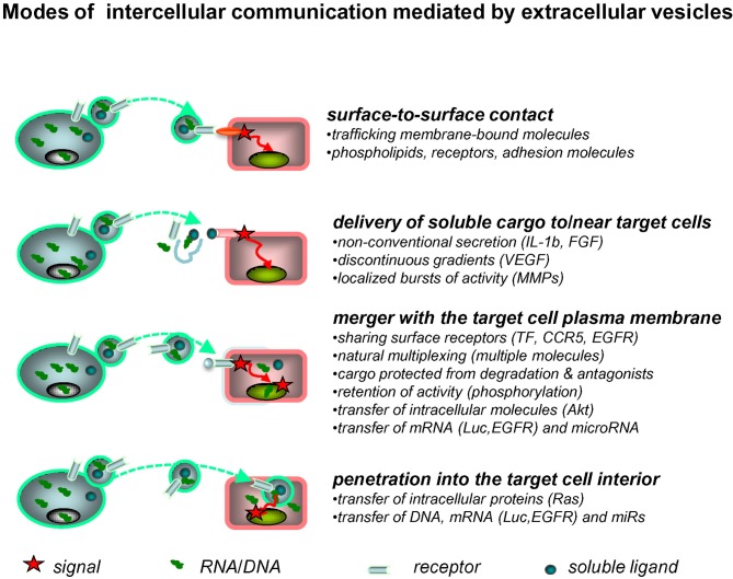
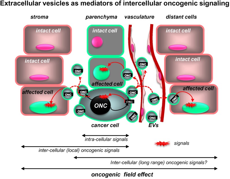
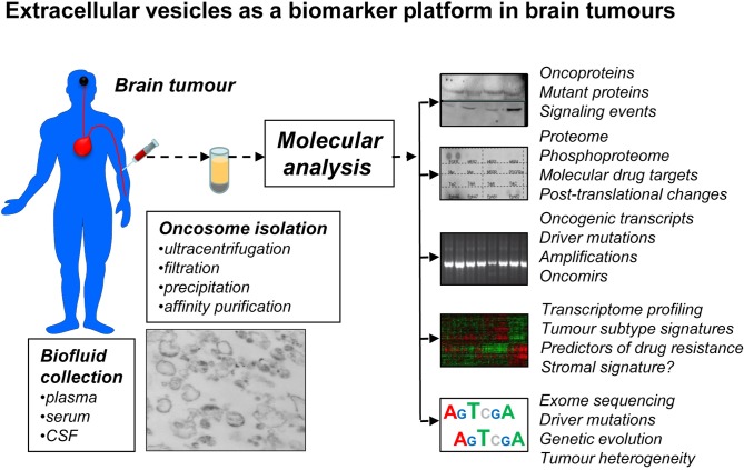
The brain is a frequent site of neoplastic growth, including both primary and metastatic tumors. The clinical intractability of many brain tumors and their distinct biology are implicitly linked to the unique microenvironment of the central nervous system (CNS) and cellular interactions within. Among the most intriguing forms of cellular interactions is that mediated by membrane-derived extracellular vesicles (EVs). Their biogenesis (vesiculation) and uptake by recipient cells serves as a unique mechanism of intercellular trafficking of complex biological messages including the exchange of molecules that cannot be released through classical secretory pathways, or that are prone to extracellular degradation. Tumor cells produce EVs containing molecular effectors of several cancer-related processes such as growth, invasion, drug resistance, angiogenesis, and coagulopathy. Notably, tumor-derived EVs (oncosomes) also contain oncogenic proteins, transcripts, DNA, and microRNA (miR). Uptake of this material may change properties of the recipient cells and impact the tumor microenvironment. Examples of transformation-related molecules found in the cargo of tumor-derived EVs include the oncogenic epidermal growth factor receptor (EGFRvIII), tumor suppressors (PTEN), and oncomirs (miR-520g). It is postulated that EVs circulating in blood or cerebrospinal fluid (CSF) of brain tumor patients may be used to decipher molecular features (mutations) of the underlying malignancy, reflect responses to therapy, or molecular subtypes of primary brain tumors [e.g., glioma or medulloblastoma (MB)]. It is possible that metastases to the brain may also emit EVs with clinically relevant oncogenic signatures. Thus, EVs emerge as a novel and functionally important vehicle of intercellular communication that can mediate multiple biological effects. In addition, they provide a unique platform to develop molecular biomarkers in brain malignancies.

Keywords: brain; cancer; exosomes; extracellular vesicles; oncogenes.


Figures

Figure 1
Figure 1
Extracellular vesicles as mediators of intercellular communication. Exchange of molecular information between cells may be mediated by EVs in several ways. Thus, surface receptors of EVs may interact directly with counter-receptors on the surface of a target cell. The latter may also come into contact with the bioactive inner cargo of EVs upon their pericellular rupture. EVs may also merge with the plasma membrane of the target cell, or penetrate into its interior via endocytosis, or other processes, to release their content of proteins and nucleic acids into the intracellular compartments (see text for details).
Figure 2
Figure 2
Extracellular vesicles as putative mediators of the intercellular propagation of oncogenic signaling. While intracellular signaling pathways elicited by mutant oncogenes (ONC) are increasingly well understood, oncoproteins (ONC) may also operate outside of the confines of cancer cells due to intercellular trafficking of their containing EVs (oncosomes). Uptake of this material by proximal non-transformed cells and at distant organ sites may trigger downstream oncogenic signals in these recipient cells and alter their phenotype and behavior. Thus, intercellular trafficking of oncoproteins (and nucleic acids) extends the range of oncogenic signaling beyond the boundaries of cells harboring the original mutation (see text for details).
Figure 3
Figure 3
Extracellular vesicles as a prospective biomarker platform in molecular diagnosis of primary and secondary brain tumors. EVs that circulate in peripheral blood or cerebrospinal fluid (CSF) can be readily recovered using several existing methods and various microfluidic and nanotechnology platforms under development. It may be possible to enrich for tumor-derived EVs (oncosomes) using tumor markers (EGFRvIII) and/or specific immunoaffinity techniques. The cargo of oncosomes may be dissected for individual molecules (oncoproteins), their activation, posttranslational processing (phosphorylation) and combinations, including for the purpose of monitoring putative drug targets and their responses to targeted therapies. EVs may provide information as to new, or pre-existing mutations that may occur in brain tumor cells, as well as scope and phylogeny of driver events, for example by sequencing nucleic acids (DNA, RNA) in the cargo. Profiling of proteins and nucleic acids may reveal signatures of brain tumor subgroups and individual variations in gene expression. The challenge is to develop technologies that would ensure sensitivity, specificity, reproducibility and processing of this information for clinical purposes.

References

    1. Abusamra A. J., Zhong Z., Zheng X., Li M., Ichim T. E., Chin J. L., Min W. P. (2005). Tumor exosomes expressing Fas ligand mediate CD8+ T-cell apoptosis. Blood Cells Mol. Dis. 35, 169–173 10.1016/j.bcmd.2005.07.001 - DOI - PubMed
    1. Aharon A., Brenner B. (2010). The role of breast cancer cells microparticles in thrombogenicity following chemotherapy. Thromb. Res. 125(Suppl. 2), S179
    1. Al-Nedawi K., Meehan B., Kerbel R. S., Allison A. C., Rak J. (2009a). Endothelial expression of autocrine VEGF upon the uptake of tumor-derived microvesicles containing oncogenic EGFR. Proc. Natl. Acad. Sci. U.S.A. 106, 3794–3799 10.1073/pnas.0804543106 - DOI - PMC - PubMed
    1. Al-Nedawi K., Meehan B., Rak J. (2009b). Microvesicles: messengers and mediators of tumor progression. Cell Cycle 8, 2014–2018 10.4161/cc.8.13.8988 - DOI - PubMed
    1. Al-Nedawi K., Meehan B., Micaleff J., Guha A., Rak J. (2010). Phosphoproteome of tumour derived microvesicles as a source of biomarkers to monitor the effects of targeted agents in glioblastoma, in Society of Neurooncology, Annual Meeting, (Montreal, QC: ).