. 2012:2012:623139.
doi: 10.5402/2012/623139. Epub 2012 Aug 12.

Designing Paclitaxel drug delivery systems aimed at improved patient outcomes: current status and challenges

Affiliations

Designing Paclitaxel drug delivery systems aimed at improved patient outcomes: current status and challenges

Madhu S Surapaneni et al. ISRN Pharmacol. 2012.

Abstract

Paclitaxel is one of the most widely used and effective antineoplastic agents derived from natural sources. It has a wide spectrum of antitumor activity, particularly against ovarian cancer, breast cancer, nonsmall cell lung cancer, head and neck tumors, Kaposi's sarcoma, and urologic malignancies. It is a highly lipophilic compound with a log P value of 3.96 and very poor aqueous solubility of less than 0.01 mg/mL. In addition, the compound lacks functional groups that are ionizable which could potentially lead to an increase in its solubility with the alteration in pH. Therefore, the delivery of paclitaxel is associated with substantial challenges. Until the introduction of Abraxane, only commercial formulation was solution of paclitaxel in cremophor, which caused severe side effects. However, in recent years, a number of approaches have been reported to solubilize paclitaxel using cosolvents and inclusion complexes. In addition, innovative approaches have been reported for passive targeting of tumors using nanoparticles, nanosuspensions, liposomes, emulsions, micelles, implants, pastes and gels. All approaches for delivery of improved therapeutic outcome have been discussed in this paper.


Figures

Figure 1
Figure 1
Structure of Paclitaxel (2α,4α,5β,7β,10β,13α)-4,10-bis(acetyloxy)-13-{[(2R,3S)-3-(benzoylamino)-2-hydroxy-3-phenylpropanoyl]oxy}-1,7-dihydroxy-9-oxo-5,20-epoxytax-11-en-2-yl benzoate.
Figure 2
Figure 2
Taxane ring.
Figure 3
Figure 3
Cumulative in vitro release of paclitaxel from PLGA-Nps (Resomer RG 502). PLGA-Nps containing paclitaxel 1% (w/w) were diluted in PBS, incubated at 37°C, and shaken horizontally. At preselected time intervals, the released drug was separated by ultracentrifugation and the residual amount of paclitaxel present in the nanospheres was determined by HPLC. (reproduced with permission from Fonseca et al., [33]).
Figure 4
Figure 4
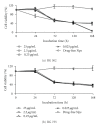
Viability of NCI-H69 cells incubated with drug-free Nps or Ptx-PLGA-Nps prepared with the RG 502 copolymer (a) or with RG 755 (b). Cells were seeded into 96-well plates and incubated with different concentrations of both formulations for 24, 72, 120 or 168 h as described (reproduced with permission from Fonseca et al., [33]).
Figure 5
Figure 5
In vitro release curves of paclitaxel loaded nanoparticles prepared under various experiment parameters (a) E5 : PLA, E7 : PLGA (75 : 25), E8 : PLGA (50 : 50); (b) ratio for PLGA-TPGS : M1 (2 : 1), M2 (1 : 1), M3 (1 : 2); (c) PLGA (75 : 25) concentration—E7: 0.125, E13: 0.188, E14: 0.25; (d) PLGA (50 : 50) concentration—E8: 0.125, E15: 0.188, E16: 0.25 (reproduced with permission from Mu and Feng, [34]).
Figure 6
Figure 6
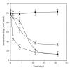
Release of paclitaxel from conventional (squares) and PEGylated (triangles) liposomes in storage conditions at 4°C (solid symbols), or in human plasma at 37°C (outline symbols) (reproduced with permission from Crosasso et al., [51]).
Figure 7
Figure 7
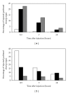
(a) Biodistribution in Balb/c mice in plasma (white), liver (black), and spleen (grey) 0.5, 3, and 24 hrs after injection of conventional paclitaxel liposomes. Standard deviations were below 5% of the mean values (reproduced with permission from Crosasso et al., [51]). (b) Biodistribution in Balb/c mice in plasma (white), liver (black), and spleen (grey) 0.5, 3 and 24 hrs after injection of PEGylated paclitaxel liposomes. Standard deviations were below 5% of the mean values (reproduced with permission from Crosasso et al., [51]).
Figure 8
Figure 8
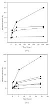
(a) Paclitaxel release at 4°C from 2′-PEG-paclitaxel (circles), 2′-succinyl-paclitaxel (triangles pointing up) and 2′-MPA-paclitaxel (triangles pointing down) in PBS buffer pH 5.8, 2′-PEG-paclitaxel (squares), 2′-succinyl-paclitaxel (triangles pointing up) and 2′-MPA-paclitaxel (triangles pointing down) in PBS buffer pH 7.4 (reproduced with permission from Ceruti et al., [52]). (b) Paclitaxel release at 37°C from 2′-PEG-paclitaxel (circles), 2′-succinyl-paclitaxel (triangles pointing down) and 2′-MPA-paclitaxel (hexagons) in PBS buffer pH 7.4, 2′-PEG-paclitaxel (squares), 2′-succinyl-paclitaxel (triangles pointing up) and 2′-MPA-paclitaxel (diamonds) in PBS buffer pH 5.8 (reproduced with permission from Ceruti et al., [52]).
Figure 9
Figure 9
In these formulations the PG : PC ratio was varied. open squares: PC only; filled squares: PG : PC (1 : 9); open circles: PG : PC (3 : 7); filled circles: PG : PC (5 : 5); open triangles: PG : PC (7 : 3); filled triangles: PG only.(reproduced with permission from Sharma and Straubinger, [53]).
Figure 10
Figure 10
Survival rate of HeLa S-3 tumor cells exposing to emulsions and Diluent 12 with or without paclitaxel. ● represents the formulated emulsion with paclitaxel, ■ represents Diluent 12 containing 6 mg paclitaxel in 1 mL 50% ethanol and 50% Cremophor EL, respectively, ○ represents emulsion without paclitaxel, and □ represents Diluent 12 without paclitaxel (reproduced with permission from Kan et al., [61]).
Figure 11
Figure 11
Survival ratio of ascitic tumor bearing mice. □ represents untreated control, ■ represents the group treated with 60 mg/kg paclitaxel emulsion, and ● represents the group treated with same quantity of paclitaxel free emulsion vehicle (reproduced with permission from Kan et al., [61]).
Figure 12
Figure 12
Comparison of treatment of Diluent 12 and emulsion on the ascitic tumor bearing mice. □ represents untreated control, ▴ represents group treated with 30 mg/kg emulsion, and ● represents group treated with Diluent 12 (reproduced with permission from Kan et al., [61]).
Figure 13
Figure 13
Mean plasma concentration of paclitaxel after IV administration of paclitaxel injection (▪) and paclitaxel microemulsion (○) in rats (n = 5) (reproduced with permission from He et al. [9]).
Figure 14
Figure 14
Inhibition of HeLa cell proliferation by paclitaxel oleate in lipid emulsions. Incubation times were 24 (●), 48 (▿), 72 (●), and 96 h ().The effect of paclitaxel in Cremophor EL/ethanol at 48 h (▴) is shown as comparison. Control growth = 100. Values are mean ± S.D., n = 4 (reproduced with permission from Lundberg et al., [64]).
Figure 15
Figure 15
Drug concentration versus time curves of [3H]paclitaxel in lipid emulsion (●), [3H]paclitaxel in Cremophor EL : ethanol 1 : 1 (v/v) (▿), and [3H]paclitaxel oleate in lipid emulsion (▪) following single i.v. bolus. Values are mean ± S.D., n = 3. *P < 0.05 versus paclitaxel in Cremophor EL/ethanol (reproduced with permission from Lundberg et al., [64]).
Figure 16
Figure 16
Time courses of paclitaxel levels in tumor of murine B16 melanoma-induced mice after i.v. administration of 50 mg/kg dose of Genexol-PM () and 20 mg/kg dose of Taxol (). Each point represents the mean ± S.D. of four mice per time point (reproduced with permission from Kim et al., [3]).
Figure 17
Figure 17
(a) Effect of PC/BS (phosphatidylcholine/bile salt) molar ratio on the solubilization potential of mixed micelles of taxol (reproduced with permission from Alkan-Onyuksel et al., [23]). (b) Effect of total lipid on the solubilization potential of mixed micelles of taxol (Reproduced with permission from Alkan-Onyuksel et al., [23])

References

    1. Mastropaolo D, Camerman A, Luo Y, Brayer GD, Camerman N. Crystal and molecular structure of paclitaxel (taxol) Proceedings of the National Academy of Sciences of the United States of America. 1995;92(15):6920–6924. - PMC - PubMed
    1. Farina V. The Chemistry and Pharmacology of Taxol and Its Derivatives. Elsevier Science; 1995.
    1. Kim SC, Kim DW, Shim YH, et al. In vivo evaluation of polymeric micellar paclitaxel formulation: toxicity and efficacy. Journal of Controlled Release. 2001;72(1–3):191–202. - PubMed
    1. Singla AK, Garg A, Aggarwal D. Paclitaxel and its formulations. International Journal of Pharmaceutics. 2002;235(1-2):179–192. - PubMed
    1. McGuire WP, Rowinsky EK, Rosenhein NB, et al. Taxol: a unique antineoplastic agent with significant activity in advanced ovarian epithelial neoplasms. Annals of Internal Medicine. 1989;111(4):273–279. - PubMed

LinkOut - more resources