. 2012 Nov;159(3):299-310.
doi: 10.1111/bjh.12033. Epub 2012 Aug 30.

Phage display generation of a novel human anti-CD1A monoclonal antibody with potent cytolytic activity

Affiliations

Phage display generation of a novel human anti-CD1A monoclonal antibody with potent cytolytic activity

Gitanjali I Bechan et al. Br J Haematol. 2012 Nov.

Abstract

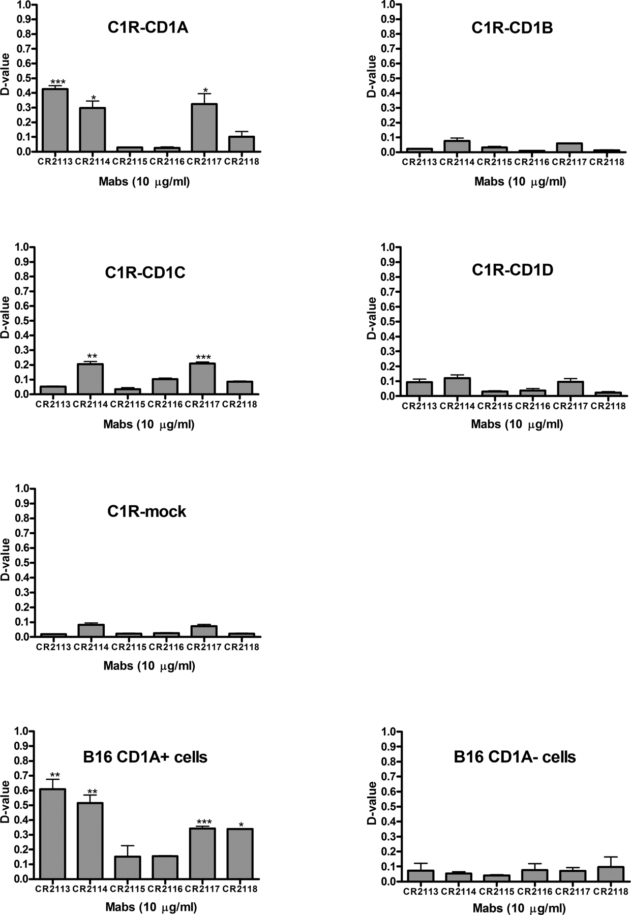
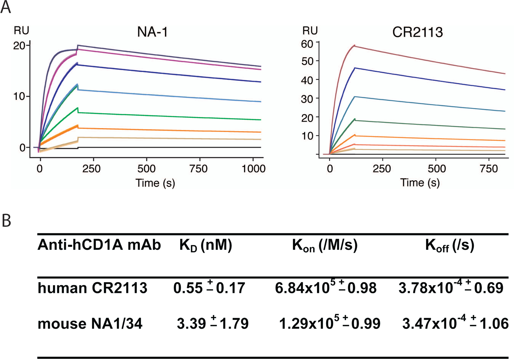
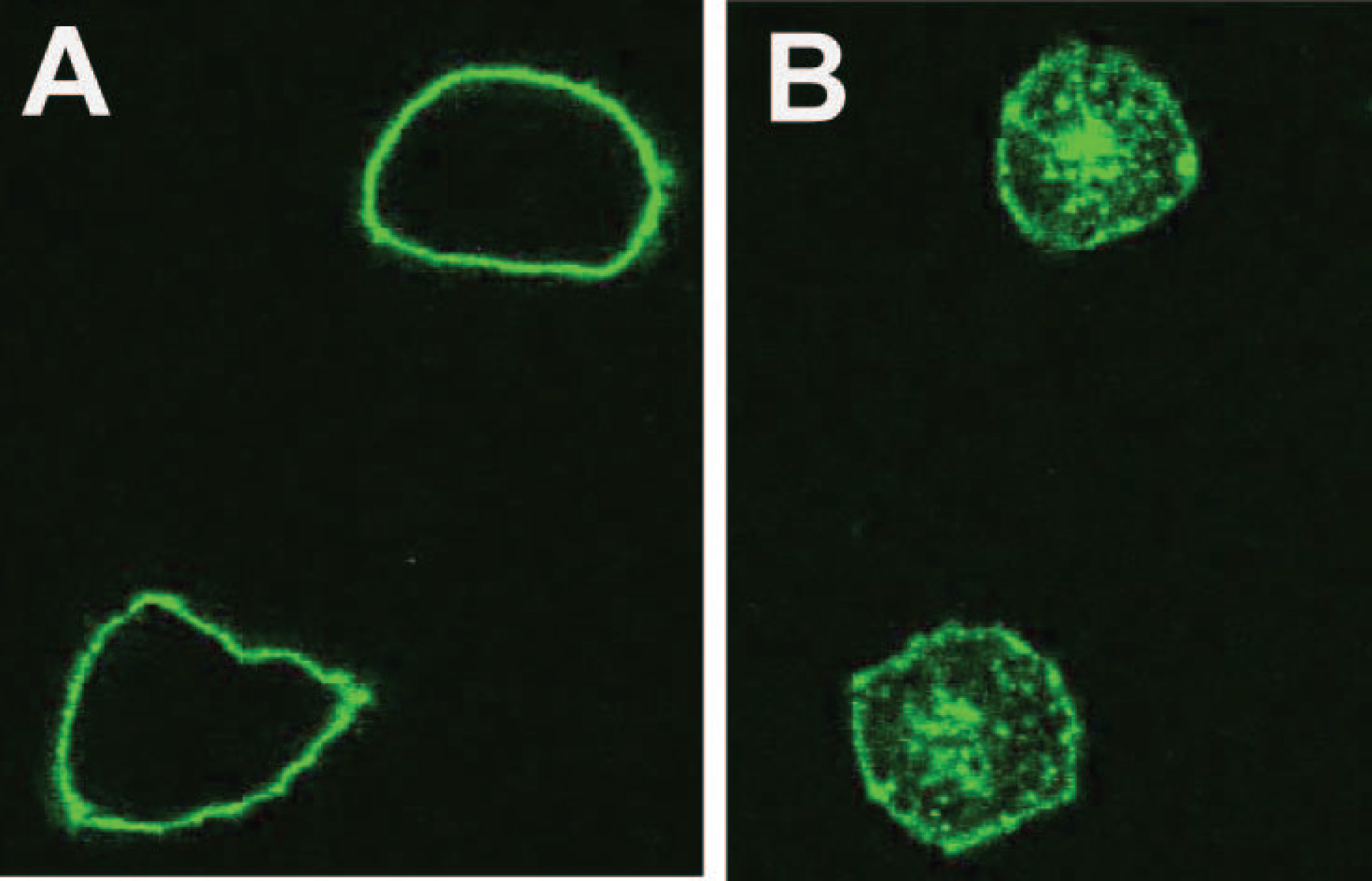
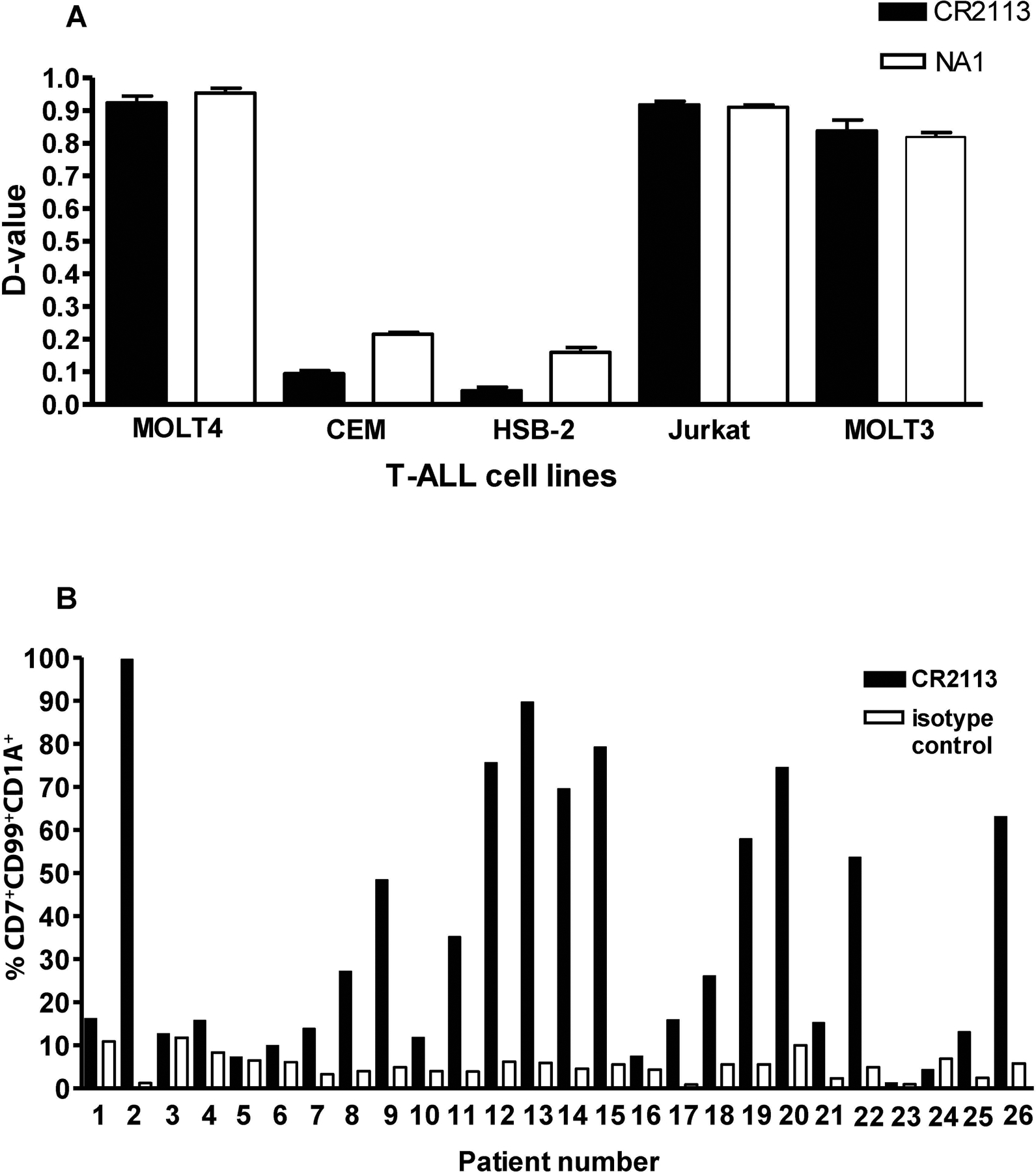
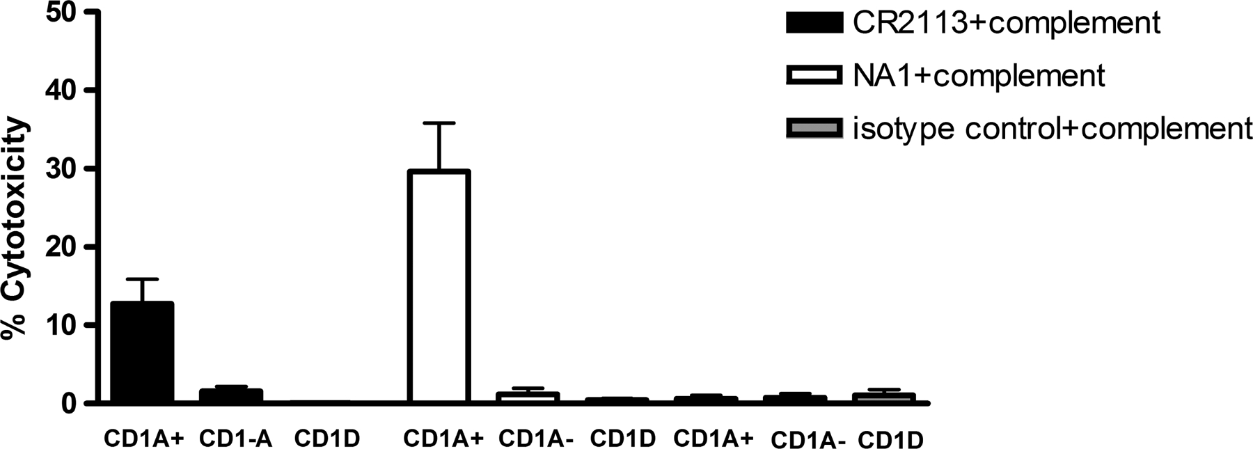
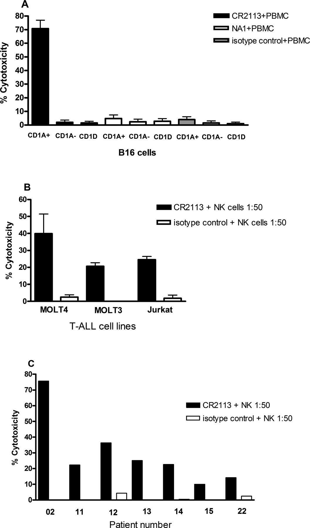
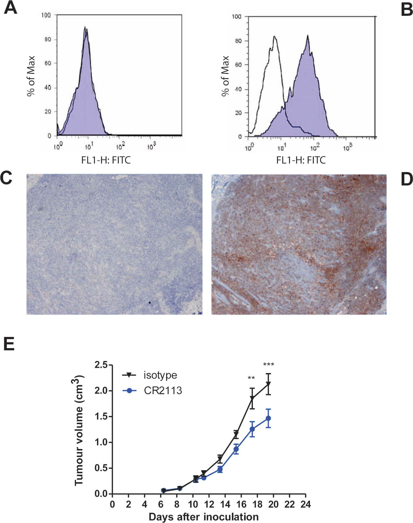
CD1A is a cell surface protein expressed on Langerhans cells and cortical thymocytes that could potentially be used as an immunotherapeutic target in Langerhans Cell Histiocytosis (LCH), the cortical subtype of T-cell acute lymphocytic leukaemia (T-ALL) and other CD1A-positive tumours. The monoclonal antibody (mAb) CR2113 was selected from a panel of six fully human mAbs isolated from a semi-synthetic phage display library, based on specificity and avidity against cells expressing CD1 antigen variants. CR2113 recognized CD1A in T-ALL cell lines and patient samples. Confocal microscopy revealed that the CR2113-CD1A complex was internalized at 37°C. Furthermore, while CR2113 induced moderate complement-dependent cytotoxicity (CDC), potent antibody-dependent cell cytotoxicity (ADCC) activity was observed against CD1A expressing cell lines as well as T-ALL cell lines and T-ALL patient samples. In vivo experiments showed that CR2113 as a naked antibody has modest but specific anti-tumour activity against CD1A-expressing tumours. CR2113 is a high-affinity human anti-CD1A mAb with significant ADCC activity. These properties make CR2113 a candidate for clinical diagnostic imaging and therapeutic targeting of LCH as well as potential use in other clinical applications.


Conflict of interest statement

Conflicts of Interest. Authors do not have any conflicts of interest. Marja van Meijer, and A.M. Kruisbeek are employees of Crucell, NV, The Netherlands. While some of the work involved in this manuscript was being done, Mark Throsby and Wilfred. Germeraad, were employees of Crucell, NV.

Figures

Figure 1.
Figure 1.
Reactivity and specificity of the human anti-CD1A monoclonal antibodies (mAbs) on different CD1 antigens expressed in the C1R and B16 cell lines. C1R cells expressing either CD1A, CD1B, CD1C or CD1D or no CD1 antigens (termed C1R mock) as well as B16 cells transfected with a viral expression vector containing the cDNA encoding CD1A or empty vector were stained with either CR2113, CR2114, CR2115, CR2116, CR2117 or CR2118 mAbs at 1, 3, and 10 μg/ml. Data for 10 μg mAb/ml are shown and representative of all three doses. Mouse IgG1, κ-FITC was used as a secondary antibody. Cells were analysed by flow cytometry using Kolmogorov-Smirnov statistics to quantitate binding as described in Methods. Specificity was determined by the level of binding to cells expressing specific CD1 isoforms compared to background, isotype control antibody staining. Statistical significance was determined by t-test (unpaired, two-tailed) analysis when comparing the binding of specific monoclonal antibodies to background staining assessed by antibody isotype control staining on cells expressing specific CD1 isoforms. Error bars represent standard deviations. CR2113, CR2114, CR2117 and CR2118 mAbs showed significant binding to CD1A expressing cells with CR2113 having the strongest specific binding characteristics. CR2114 and CR2117 also showed some affinity for CD1C. Legend: ***: p< 0.001, **: p< 0.01, *: p<0.05, ns: p>0.05
Figure 2.
Figure 2.
Affinity and kinetic binding measurements by surface plasmon resonance. Binding kinetics of mouse and human anti-human CD1A mAbs NA1/34 and CR2113, respectively, were determined by surface plasmon resonance. Sensograms of mouse mAb NA1/34 (left panel) and human CR2113 (right panel) are depicted (A). Increasing concentrations of hCD1A from 0.5–32nM (CR2113) and 10–640 nM (NA1/34) in two-fold dilutions were injected and the binding responses were superimposed. A representative experiment is shown. Affinity and kinetic parameters are summarized in B. Numbers are mean (± s.e.m) of two (NA1/34) and three (CR2113) separate experiments.
Figure 3.
Figure 3.
Internalization of AF488-CR2113. B16 CD1A+ cells were incubated at either 4°C (left) or 37°C (right) with CR2113-AF488for 1 h. Cells kept at 4°C during incubation showed only surface binding of CR2113-AF488. Internalization of the CD1A-CR2113-AF488 complex at 37°C was evident from capping and punctate staining of endocytic vessels. AF488 labelled isotype control antibody did not show binding or internalization on CD1A-positive or -negative cells and CR2113-AF488 did not show binding or internalization on CD1A-negative cells (data not shown).
Figure 4.
Figure 4.
CD1A expression in leukaemia cell lines and patient samples. Five T-ALL cell lines and 26 T-ALL patient samples were stained with CR2113 and screened for CD1A expression by flow cytometry. CR2113 detected CD1A expression in 3 of 5 T-ALL cell lines (A) and 9 of 26 T-ALL patient samples (B). Panel B also shows the percentage of CD7+/CD99+/CD1A+ cells detected for each of the leukaemia samples compared to isotype control mAb.
Figure 5.
Figure 5.
Complement-Dependent Cytotoxicty. B16 cells expressing CD1A, CD1D or B16 cells transfected with vector only were incubated with CR2113, NA1/34 or isotype control alone or with human serum for 5 h at 37°C. CR2113 induced a moderate level while NA1/34 produced a higher cytotoxicity level of CDC in CD1A+ cells.
Figure 6.
Figure 6.
Antibody-Dependent Cell Cytotoxicity. B16 cells expressing CD1A, CD1D or B16 cells transfected with vector only were incubated with CR2113, NA1/34 or isotype control alone or with either peripheral blood mononuclear cells (PBMC) or natural killer (NK) cells for 5 hs at 37°C. CR2113 induced a high level of ADCC in the CD1A+ cells (A). NA1/34 was not effective in inducing cytotoxicity by ADCC. ADCC experiments with CR2113 in combination with NK cells (1:50 ratio) were also performed on the CD1A positive cell lines and patient samples. CR2113 induced ADCC in 3 of 3 CD1A+ T-ALL cell lines (B) and in 7 of 9 CD1A+ T-ALL patient samples (C).
Figure 7.
Figure 7.
Engrafted B16 mouse melanoma tumours retain CD1A expression by flow cytometry and immunohistochemistry. (A, B) By flow cytometry, harvested B16 melanoma tumours derived from CD1A-B16 melanoma cells do not stain with mAb CR2113 anti-human CD1A antibody (A) while CD1A+ B16 melanoma cells stain positive with mAb CR2113 anti-human CD1A antibody (B). The transparent curve represents isotype control mAb (superimposed over blue curve in panel A) and the blue curve is staining with mAb CR2113. (C, D) B16 melanoma tumours derived from CD1A+ B16 melanoma cells did not show positive staining with isotype control antibody followed by staining with Haematoxylin (C) while CD1A+ B16 melanoma tumours stain showed strong staining with an anti-CD1A antibody (Becton Dickinson, Franklin Lakes, NJ) (D). (E) Efficacy of mAb CR2113 antibody against B16 mouse melanoma tumours expressing CD1A antigen. NOD/SCID mice were subcutaneously injected with 1×10⁶ tumour cells in each flank while they were under anesthesia to assure consistent site and injection characteristics. Tumours grew at a uniform rate. Animals were treated with mAb CR2113 (Blue) or isotype control antibody (Black) on days 2, 6, 10, 14 and 18 as described in the text. Graphs represent the average tumour volume over time for all animals in a particular arm of the experiment. Error bars represent the standard error of the mean. Two–way ANOVA identifies statistical significance of p<0.001 (***) as indicated in the figure. Of note, anti-CD1A CR2113 did not have an inhibitory effect on non-CD1A expressing B16 derived tumours (data not shown).

References

    1. Adams GP & Weiner LM (2005) Monoclonal antibody therapy of cancer. Nat Biotechnol, 23, 1147–1157. - PubMed
    1. Allam JP & Novak N (2006) The pathophysiology of atopic eczema. Clinical and Experimental Dermatology, 31, 89–93. - PubMed
    1. Allen CE, Li L, Peters TL, Leung HC, Yu A, Man TK, Gurusiddappa S, Phillips MT, Hicks MJ, Gaikwad A, Merad M & McClain KL (2010) Cell-specific gene expression in Langerhans cell histiocytosis lesions reveals a distinct profile compared with epidermal Langerhans cells. Journal of Immunology, 184, 4557–4567. - PMC - PubMed
    1. Amiot M, Bernard A, Raynal B, Knapp W, Deschildre C & Boumsell L (1986) Heterogeneity of the first cluster of differentiation: characterization and epitopic mapping of three CD1 molecules on normal human thymus cells. Journal of Immunology, 136, 1752–1758. - PubMed
    1. Amiot M, Dastot H, Degos L, Schmid M & Boumsell L (1987) ‘Leukocyte Typing III’. Oxford Univ Press, Oxford.

Publication types

MeSH terms