. 2012 Aug 30:9:207.
doi: 10.1186/1742-2094-9-207.

Protein expression profiling of inflammatory mediators in human temporal lobe epilepsy reveals co-activation of multiple chemokines and cytokines

Affiliations

Protein expression profiling of inflammatory mediators in human temporal lobe epilepsy reveals co-activation of multiple chemokines and cytokines

Anne A Kan et al. J Neuroinflammation. .

Abstract

Mesial temporal lobe epilepsy (mTLE) is a chronic and often treatment-refractory brain disorder characterized by recurrent seizures originating from the hippocampus. The pathogenic mechanisms underlying mTLE remain largely unknown. Recent clinical and experimental evidence supports a role of various inflammatory mediators in mTLE. Here, we performed protein expression profiling of 40 inflammatory mediators in surgical resection material from mTLE patients with and without hippocampal sclerosis, and autopsy controls using a multiplex bead-based immunoassay. In mTLE patients we identified 21 upregulated inflammatory mediators, including 10 cytokines and 7 chemokines. Many of these upregulated mediators have not previously been implicated in mTLE (for example, CCL22, IL-7 and IL-25). Comparing the three patient groups, two main hippocampal expression patterns could be distinguished, pattern I (for example, IL-10 and IL-25) showing increased expression in mTLE + HS patients compared to mTLE-HS and controls, and pattern II (for example, CCL4 and IL-7) showing increased expression in both mTLE groups compared to controls. Upregulation of a subset of inflammatory mediators (for example, IL-25 and IL-7) could not only be detected in the hippocampus of mTLE patients, but also in the neocortex. Principle component analysis was used to cluster the inflammatory mediators into several components. Follow-up analyses of the identified components revealed that the three patient groups could be discriminated based on their unique expression profiles. Immunocytochemistry showed that IL-25 IR (pattern I) and CCL4 IR (pattern II) were localized in astrocytes and microglia, whereas IL-25 IR was also detected in neurons. Our data shows co-activation of multiple inflammatory mediators in hippocampus and neocortex of mTLE patients, indicating activation of multiple pro- and anti-epileptogenic immune pathways in this disease.


Figures
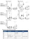
Figure 1
Figure 1
HC and CX expression patterns of inflammatory proteins in mTLE. Scatter plots of protein levels per individual patient, per group. Horizontal lines represent the median expression level of the group. Post-hoc analysis of the data revealed three expression patterns among the patient groups. We classified them as Type I to III (I: A and B, II: C and D, III: E). Additionally, different patterns were detected depending on the type of tissue, hence we classified an up-regulation in HC only as A (A and C) and an up-regulation in both HC and CX as B (B and D). * indicates significant difference. All significantly changed proteins showed one of these three expression patterns (G) (see Figure 2)
Figure 2
Figure 2
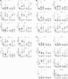
HC and CX expression patterns of other inflammatory proteins. Interleukins (A), chemokines (B), miscellaneous inflammatory proteins (C), and inflammatory proteins with p-values 0.0017-0.05 which were not tested post-hoc (D). Scatterplots of individual protein levels per patient group. Horizontal lines represent the median expression level of the group
Figure 3
Figure 3
Hippocampal expression patterns of IL-25 and CCL4 in Control, mTLE-HS and mTLE + HS patients. Photomicrographs showing typical examples of Il-25 and CCL4 staining in the hippocampal CA1 region (A) and the DG/CA4 region (B). IL-25 immunoreactivity (IR) is evident in cells with neuronal morphology in all three patient groups (A, B); note the increased IL-25 staining in the mTLE + HS hippocampus in small cells. CCL4 IR is low in controls (A, B). Increased CCL4 IR is detected in both mTLE patient groups in the DG-CA4 area (B) and in the CA1 area of mTLE + HS patients (A). Scale bar = 200 μm.The insets are higher power magnifications taken from the same anatomical area
Figure 4
Figure 4
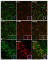
IL-25 IR in neurons, astrocytes and microglia. IFC reveals IL-25 IR (in green) co-localizing with three CNS cell type markers (red), (A) Iba1, a microglial marker, (B) GFAP, an astrocytic marker and (C) NeuN, a neuronal marker. Panel A,B: CA4; Panel C: dentate gyrus. Note that IL-25 IR is also present in the astrocytic endfeet surrounding the bloodvessels (B). The insets are higher power magnifications taken from a representative area, with the added nuclear marker DAPI (blue). Scale bar = 40 μm
Figure 5
Figure 5
CCL4 IR in astrocytes and reactive astrocytes. IFC reveals CCL4 (in green) co-localizing with two types of glial cell markers (red), (A) astrocytic marker GFAP and (B) reactive astrocytic marker Vimentin. CCL4 IR is also present in the astrocytic endfeet of the GFAP positive astrocytes (A). Pictures are taken of representative areas in the CA4 region. The insets are higher power magnifications taken from the same anatomical area, with the added nuclear marker DAPI (blue). Scale bar = 40 μm
Figure 6
Figure 6
Correlation network plots for all patient groups. Network plots depicting significant correlations between levels of inflammatory mediators in Control patients (A), mTLE -HS patients (B) and mTLE + HS patients (C). Line (edge) color indicates in which tissue correlation was found: red = HC, blue = CX, green = HC & CX. Per patient group two main non-overlapping correlation networks were identified. Note that correlation networks are remarkably similar among the patient groups. Inflammatory mediators like IL-5, IL-13 and IL-22 all display >4 correlations with other inflammatory mediators. Only proteins that were in a network of more than 6 correlations (nodes) are depicted. In addition, 5 significant correlations were found in the total data set. In Control HC IL-10 & CCL3 correlated with IL-25 and in mTLE-HS CX Cathepsin-S correlated with ICAM1. All correlations had a correlation coefficient > 0.85 with a Bonferoni corrected p-value < 0.0017
Figure 7
Figure 7
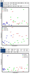
Major components identified by principle component analyses reveal pathology-specific immunological networks in the HC and the CX. PCA results for the main components identified by the analysis in the HC (A) and the CX (B). Proteins are depicted in sequence of factor loading, all depicted proteins of the components have a factor loading of >0.4 (is >16% of explained variance). Individual patient component scores were plotted against each other and revealed patient group-dependent clusters, component 1 versus 2 (A) segregated both mTLE patient groups from controls in HC (A) and CX (B), whereas the PCA plot for component 1 versus 3 in the HC segregated all three patient groups (B). C-1 to 3 = component 1 to 3

References

    1. Leonardi M, Ustun TB. The global burden of epilepsy. Epilepsia. 2002;43(Suppl 6):21–25. - PubMed
    1. Engel J Jr. A proposed diagnostic scheme for people with epileptic seizures and with epilepsy: report of the ILAE Task Force on Classification and Terminology. Epilepsia. 2001;42(6):796–803. doi: 10.1046/j.1528-1157.2001.10401.x. - DOI - PubMed
    1. Kwan P, Brodie MJ. Clinical trials of antiepileptic medications in newly diagnosed patients with epilepsy. Neurology. 2003;60:S2–S12. - PubMed
    1. Cascino GD. When drugs and surgery don’t work. Epilepsia. 2008;49(Suppl 9):79–84. - PubMed
    1. Proper EA, Oestreicher AB, Jansen GH, Veelen CW, van Rijen PC, Gispen WH, de Graan PN. Immunohistochemical characterization of mossy fibre sprouting in the hippocampus of patients with pharmaco-resistant temporal lobe epilepsy. Brain. 2000;123(Pt 1):19–30. - PubMed

Publication types

MeSH terms