Review
. 2012 Aug 31;111(6):778-99.
doi: 10.1161/CIRCRESAHA.111.256859.

Role of phospholipid oxidation products in atherosclerosis

Affiliations
Review

Role of phospholipid oxidation products in atherosclerosis

Sangderk Lee et al. Circ Res. .

Abstract

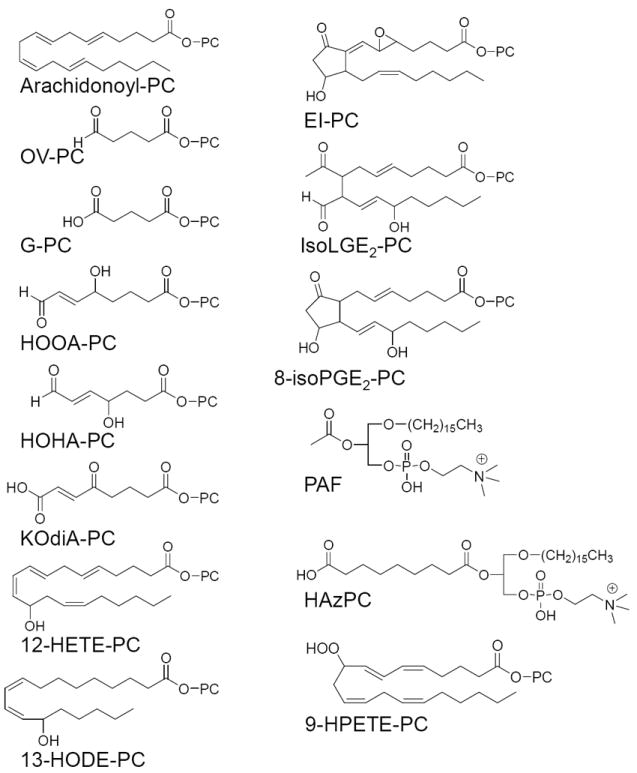
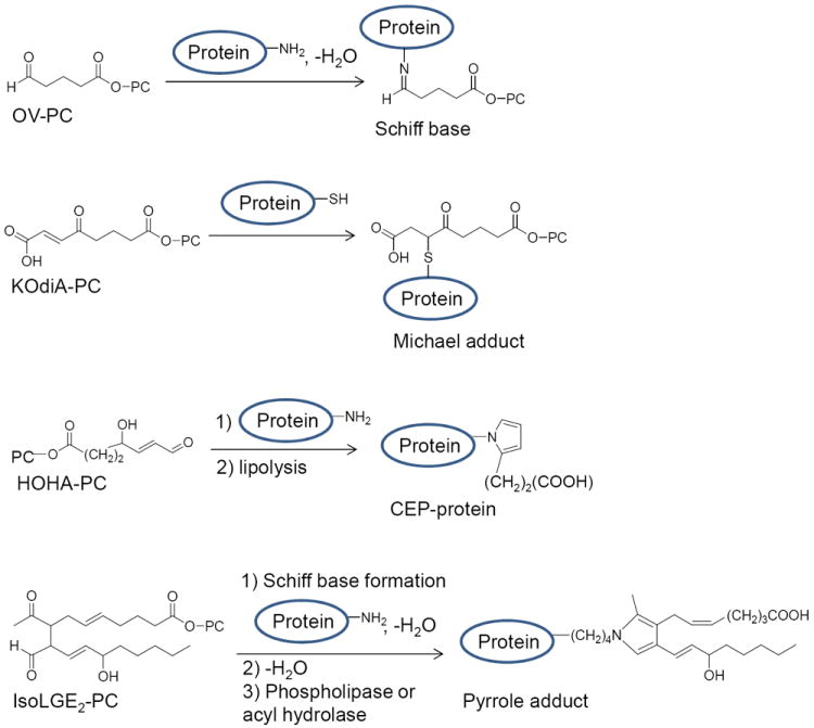
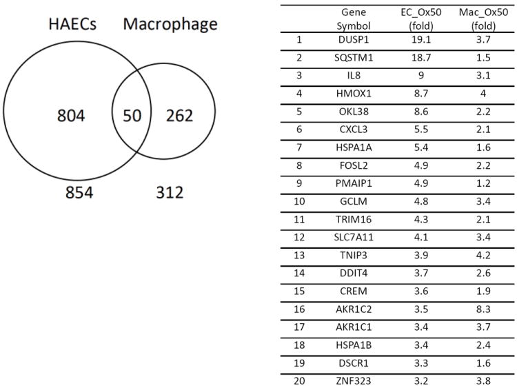
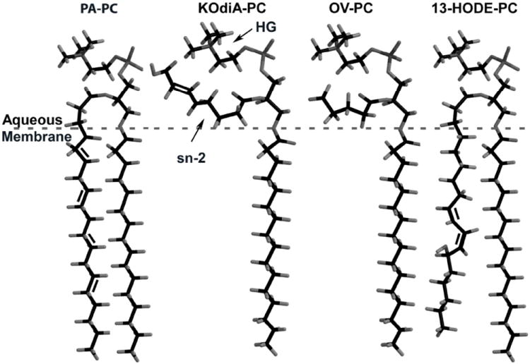
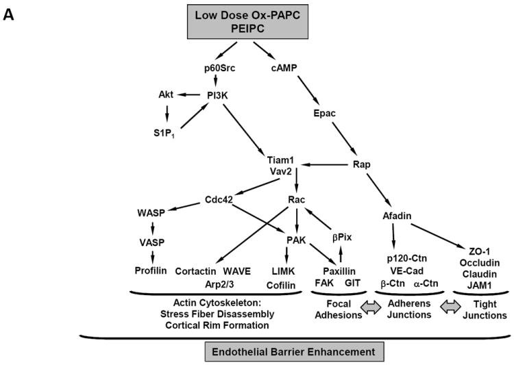
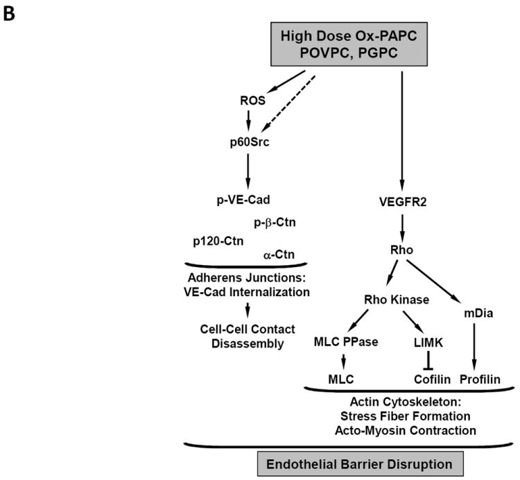
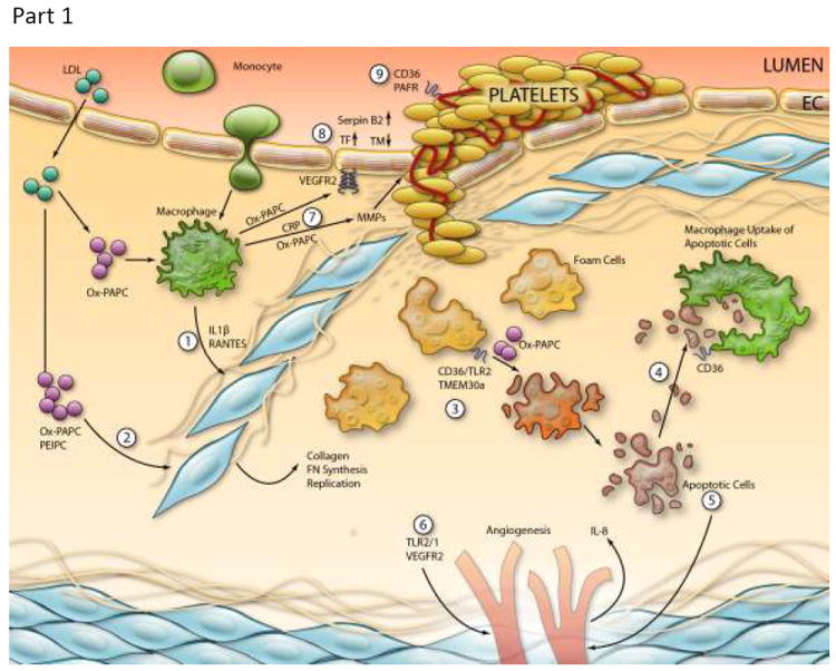
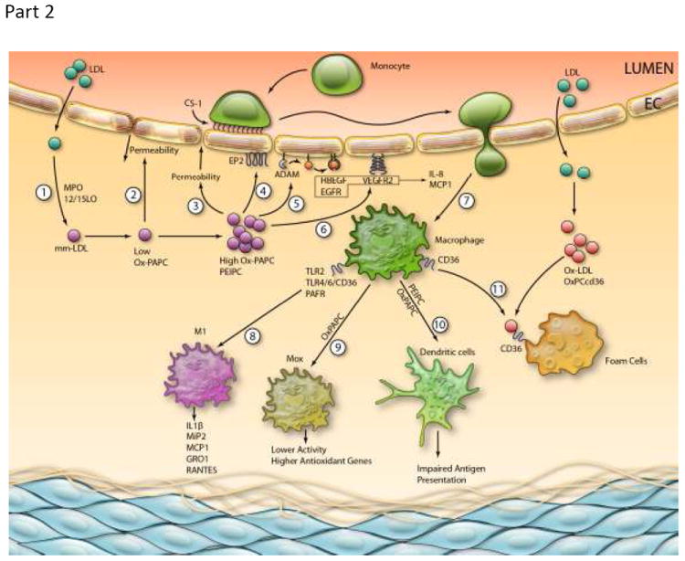
There is increasing clinical evidence that phospholipid oxidation products (Ox-PL) play a role in atherosclerosis. This review focuses on the mechanisms by which Ox-PL interact with endothelial cells, monocyte/macrophages, platelets, smooth muscle cells, and HDL to promote atherogenesis. In the past few years major progress has been made in identifying these mechanisms. It has been recognized that Ox-PL promote phenotypic changes in these cell types that have long-term consequences for the vessel wall. Individual Ox-PL responsible for specific cellular effects have been identified. A model of the configuration of bioactive truncated Ox-PL within membranes has been developed that demonstrates that the oxidized fatty acid moiety protrudes into the aqueous phase, rendering it accessible for receptor recognition. Receptors and signaling pathways for individual Ox-PL species are now determined and receptor independent signaling pathways identified. The effects of Ox-PL are mediated both by gene regulation and transcription independent processes. It has now become apparent that Ox-PL affects multiple genes and pathways, some of which are proatherogenic and some are protective. However, at concentrations that are likely present in the vessel wall in atherosclerotic lesions, the effects promote atherogenesis. There have also been new insights on enzymes that metabolize Ox-PL and the significance of these enzymes for atherosclerosis. With the knowledge we now have of the regulation and effects of Ox-PL in different vascular cell types, it should be possible to design experiments to test the role of specific Ox-PL on the development of atherosclerosis.


Figures

Figure 1
Figure 1
Ox-PL lipids. PC = 1-acyl-2-lyso-sn-glycero-3-phosphatidylcholine. Only the sn-2 position composition is shown for all Ox-PL except those forming an ether bond at the sn-1 position.
Figure 2
Figure 2
Interaction of Ox-PL lipids with proteins. Examples of three different types of interactions are shown. Only the composition of the sn-2 position is shown.
Figure 3
Figure 3
Comparison of the effect of Ox-PAPC treatment (50μg/ml for 4 hours) on gene regulation in human endothelial cells and human macrophages. A list of the top 20 genes in the overlap category is shown.
Figure 4
Figure 4
Lipid Whisker Model of the conformation of PAPC, 1-palmitoyl-2-(7-carboxy-5-oxo-hept-6E-enoyl)-sn-glycero-3-phosphocholine (KOdiA-PC), POVPC and 13-HODE-PC within membranes. An arrow points to the sn-3 and head group positions of KOdiA-PC.
Figure 5
Figure 5
A. Barrier-protecitve mechanisms stimulated by low doses of OxPAPC B. Barrier-disruptive mechanisms stimulated by high doses of OxPAPC Signal transduction pathways leading to Ox-PAPC regulation of endothelial monolayer barrier function. (A) Effects of low concentrations that strengthen the barrier.(B) Effects of higher but non- toxic concentrations that breakdown the barrier.
Figure 5
Figure 5
A. Barrier-protecitve mechanisms stimulated by low doses of OxPAPC B. Barrier-disruptive mechanisms stimulated by high doses of OxPAPC Signal transduction pathways leading to Ox-PAPC regulation of endothelial monolayer barrier function. (A) Effects of low concentrations that strengthen the barrier.(B) Effects of higher but non- toxic concentrations that breakdown the barrier.
Figure 6
Figure 6
Model of Ox-PL regulation of atherosclerosis: (A) Early Lesions (1) LDL enters the vessel wall and is oxidized by MPO and 12/15 LO to form oxidation products including Ox-PAPC. Low doses of Ox-PAPC decrease the permeability of the EC monolayer by forming adherens junctions. Higher levels of Ox-PAPC cause a strong increase in monolayer permeability due to junction breakdown and stress fiber formation, resulting in increased entry of LDL into the vessel wall. (4) Ox-PAPC binds to the E-type prostaglandin receptor (EP2) receptor, causing deposition of CS-1 Fibronectin on the apical surface which binds monocytes. (5) Ox-PAPC activates specific a dysintegrin and metalloproteinases (ADAMs) to cause release of active HBEGF and activation of epidermal growth factor receptor (EGFR), leading to IL-8 and MCP-1 synthesis. (6) Ox-PAPC also activates VEGFR2, leading to IL-8 and MCP-1 synthesis. (7) These chemokines facilitate entry of monocytes into the vessel wall. (8) Ox-PL acting on TLR2, TLR4/6/CD36 and PAFR cause some monocytes to differentiate into M1 macrophages producing chemokines. (9) Ox-PAPC causes differentiation of some macrophages into Mox, which have high levels of anti-oxidant enzymes and lower chemokine synthesis. (10) Ox-PAPC causes differentiation of some monocytes into dendritic cells with impaired presentation of lipid antigens. (11) Macrophages further oxidize LDL to form Ox-LDL. Ox-PCCD36 acting on CD36 in the presence of Ox-LDL causes foam cell formation. (B) Advanced Lesions. (1) In the presence of Ox-PAPC and PAF-like lipids, macrophages make: Interleukin 1 beta (IL-1β) and RANTES. These chemokines and a direct effect of Ox-PAPC on SMC cause migration and proliferation and matrix production of SMC. These SMC cover the foam cells that accumulate under the endothelium. The interaction of Ox-PAPC with CD36/TLR2 and with UPR activators and the interaction of PAF-like lipids with TMEM30a cause macrophage apoptosis. (4) Ox-PS/PCCD36 in the apopotic cell membrane bind to CD36 in macrophages, leading to macrophage uptake of the apoptotic cells. (5) Some apoptotic fragments stimulate EC to make IL-8, an angiogenic cytokine. (6) CEP activation of TLR2/TLR1 causing integrin activation and Ox-PAPC acting to increase VEGFA cause angiogenesis of adventitial vessels into the media and intima. (7) C-reactive protein (CRP) and Ox-PAPC interacting with CD36 stimulates macrophage production of metalloproteinase. This weakens the plaque and can lead to plaque rupture. (8) Ox-PAPC activation of VEGFR2 increases tissue factor synthesis in the endothelium. Ox-PAPC also causes and increases in Serpin B2 and a decrease in thrombomodulin. Ox-PCCD36 acting on CD36 and PAF-like lipids acting on PAFR cause increased aggregability of platelets. (Illustration Credit: Ben Smith).
Figure 6
Figure 6
Model of Ox-PL regulation of atherosclerosis: (A) Early Lesions (1) LDL enters the vessel wall and is oxidized by MPO and 12/15 LO to form oxidation products including Ox-PAPC. Low doses of Ox-PAPC decrease the permeability of the EC monolayer by forming adherens junctions. Higher levels of Ox-PAPC cause a strong increase in monolayer permeability due to junction breakdown and stress fiber formation, resulting in increased entry of LDL into the vessel wall. (4) Ox-PAPC binds to the E-type prostaglandin receptor (EP2) receptor, causing deposition of CS-1 Fibronectin on the apical surface which binds monocytes. (5) Ox-PAPC activates specific a dysintegrin and metalloproteinases (ADAMs) to cause release of active HBEGF and activation of epidermal growth factor receptor (EGFR), leading to IL-8 and MCP-1 synthesis. (6) Ox-PAPC also activates VEGFR2, leading to IL-8 and MCP-1 synthesis. (7) These chemokines facilitate entry of monocytes into the vessel wall. (8) Ox-PL acting on TLR2, TLR4/6/CD36 and PAFR cause some monocytes to differentiate into M1 macrophages producing chemokines. (9) Ox-PAPC causes differentiation of some macrophages into Mox, which have high levels of anti-oxidant enzymes and lower chemokine synthesis. (10) Ox-PAPC causes differentiation of some monocytes into dendritic cells with impaired presentation of lipid antigens. (11) Macrophages further oxidize LDL to form Ox-LDL. Ox-PCCD36 acting on CD36 in the presence of Ox-LDL causes foam cell formation. (B) Advanced Lesions. (1) In the presence of Ox-PAPC and PAF-like lipids, macrophages make: Interleukin 1 beta (IL-1β) and RANTES. These chemokines and a direct effect of Ox-PAPC on SMC cause migration and proliferation and matrix production of SMC. These SMC cover the foam cells that accumulate under the endothelium. The interaction of Ox-PAPC with CD36/TLR2 and with UPR activators and the interaction of PAF-like lipids with TMEM30a cause macrophage apoptosis. (4) Ox-PS/PCCD36 in the apopotic cell membrane bind to CD36 in macrophages, leading to macrophage uptake of the apoptotic cells. (5) Some apoptotic fragments stimulate EC to make IL-8, an angiogenic cytokine. (6) CEP activation of TLR2/TLR1 causing integrin activation and Ox-PAPC acting to increase VEGFA cause angiogenesis of adventitial vessels into the media and intima. (7) C-reactive protein (CRP) and Ox-PAPC interacting with CD36 stimulates macrophage production of metalloproteinase. This weakens the plaque and can lead to plaque rupture. (8) Ox-PAPC activation of VEGFR2 increases tissue factor synthesis in the endothelium. Ox-PAPC also causes and increases in Serpin B2 and a decrease in thrombomodulin. Ox-PCCD36 acting on CD36 and PAF-like lipids acting on PAFR cause increased aggregability of platelets. (Illustration Credit: Ben Smith).

Similar articles

Cited by

References

    1. Berliner JA, Leitinger N, Tsimikas S. The role of oxidized phospholipids in atherosclerosis. J Lipid Res. 2009;50(Suppl):S207–212. - PMC - PubMed
    1. Bochkov VN, Oskolkova OV, Birukov KG, Levonen AL, Binder CJ, Stockl J. Generation and biological activities of oxidized phospholipids. Antioxidants & redox signaling. 2010;12:1009–1059. - PMC - PubMed
    1. Hahn BH, McMahon M. Atherosclerosis and systemic lupus erythematosus: The role of altered lipids and of autoantibodies. Lupus. 2008;17:368–370. - PubMed
    1. Tsimikas S, Mallat Z, Talmud PJ, Kastelein JJ, Wareham NJ, Sandhu MS, Miller ER, Benessiano J, Tedgui A, Witztum JL, Khaw KT, Boekholdt SM. Oxidation-specific biomarkers, lipoprotein(a), and risk of fatal and nonfatal coronary events. J Am Coll Cardiol. 2010;56:946–955. - PubMed
    1. Choi SH, Chae A, Miller E, Messig M, Ntanios F, DeMaria AN, Nissen SE, Witztum JL, Tsimikas S. Relationship between biomarkers of oxidized low-density lipoprotein, statin therapy, quantitative coronary angiography, and atheroma: Volume observations from the reversal (reversal of atherosclerosis with aggressive lipid lowering) study. J Am Coll Cardiol. 2008;52:24–32. - PubMed

Publication types