. 2012 Nov;154(5):767-778.e2.
doi: 10.1016/j.ajo.2012.05.007. Epub 2012 Aug 28.

Histologic development of the human fovea from midgestation to maturity

Affiliations

Histologic development of the human fovea from midgestation to maturity

Anita Hendrickson et al. Am J Ophthalmol. 2012 Nov.

Abstract

Purpose: To describe the histologic development of the human central retina from fetal week (Fwk) 22 to 13 years.

Design: Retrospective observational case series.

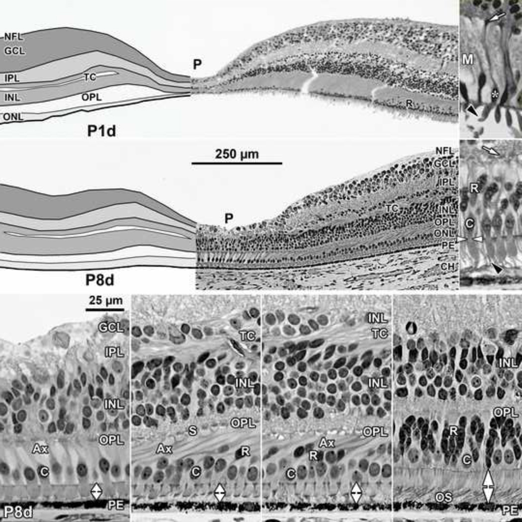
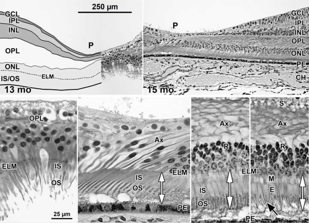
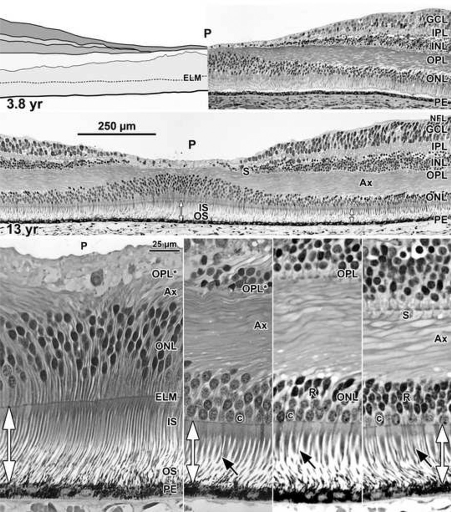
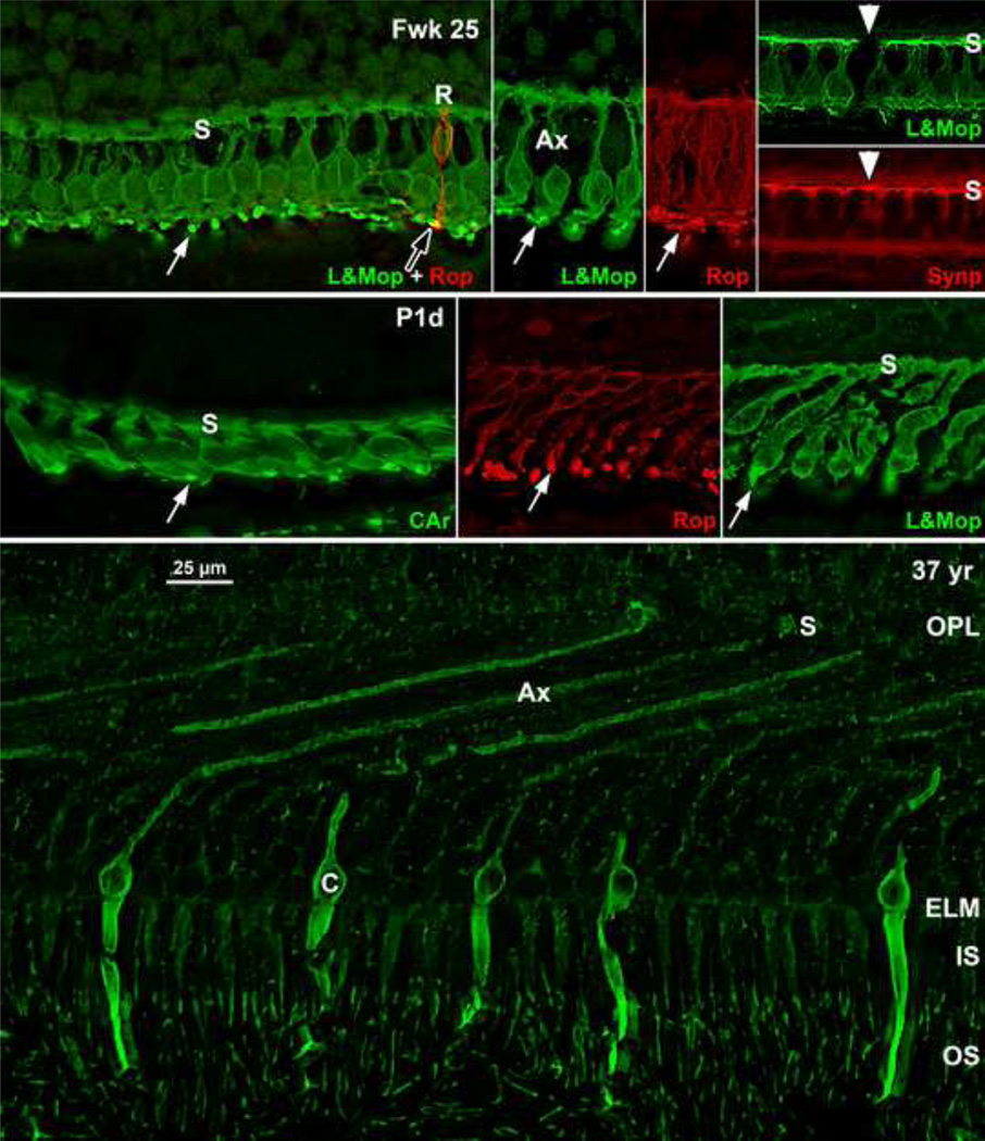
Methods: Retinal layers and neuronal substructures were delineated on foveal sections of fixed tissue stained in azure II-methylene blue and on frozen sections immunolabeled for cone, rod, or glial proteins. Postmortem tissue was from 11 eyes at Fwk 20-27; 8 eyes at Fwk 28-37; 6 eyes at postnatal 1 day to 6 weeks; 3 eyes at 9 to 15 months; and 5 eyes at 28 months to 13 years.

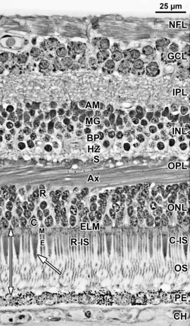
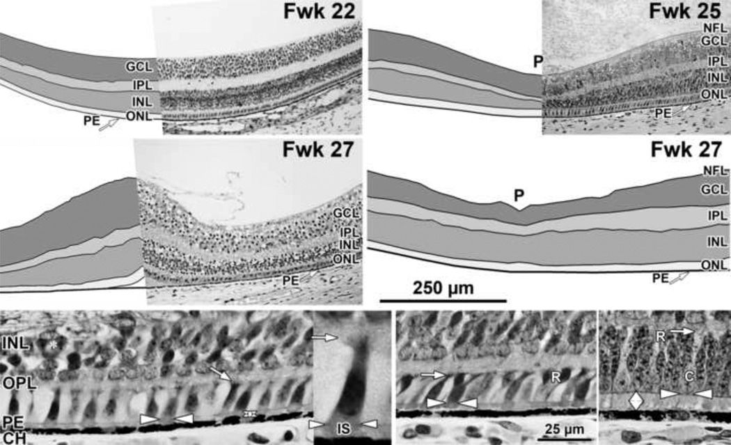
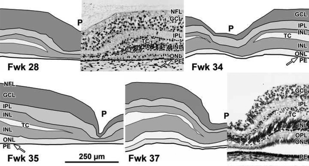
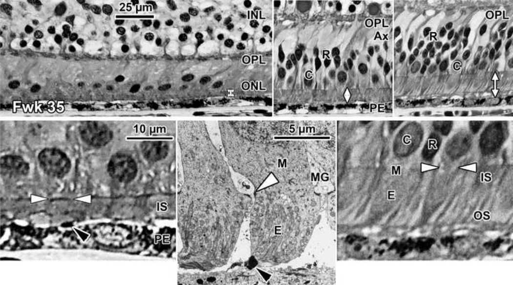
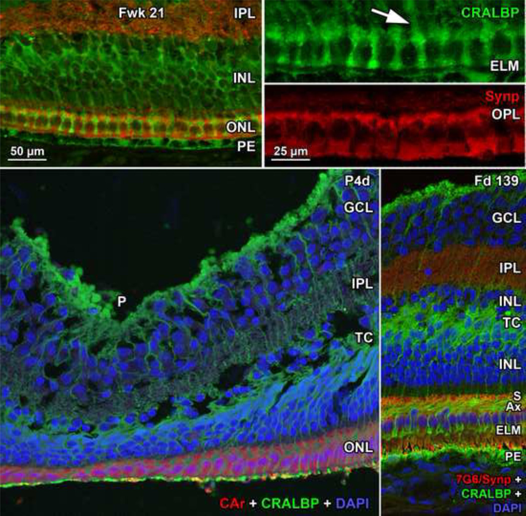
Results: At Fwk 20-22 the fovea could be identified by the presence of a single layer of cones in the outer nuclear layer. Immunolabeling detected synaptic proteins, cone and rod opsins, and Müller glial processes separating the photoreceptors. The foveal pit appeared at Fwk 25, involving progressive peripheral displacement of ganglion cell, inner plexiform, and inner nuclear layers. The pit became wider and shallower after birth, and appeared mature by 15 months. Between Fwk 25 and Fwk 38, all photoreceptors developed more distinct inner and outer segments, but these were longer on peripheral than foveal cones. After birth the foveal outer nuclear layer became much thicker as cone packing occurred. Cone packing and neuronal migration during pit formation combined to form long central photoreceptor axons, which changed the outer plexiform layer from a thin sheet of synaptic pedicles into the thickest layer in the central retina by 15 months. Foveal inner and outer segment length matched peripheral cones by 15 months and was 4 times longer by 13 years.

Conclusions: These data are necessary to understand the marked changes in human retina from late gestation to early adulthood. They provide qualitative and quantitative morphologic information required to interpret the changes in hyper- and hyporeflexive bands in pediatric spectral-domain optical coherence tomography images at the same ages.


Conflict of interest statement

Financial Disclosures:

Cynthia A. Toth receives royalties through her university from Alcon; obtained research support for other studies from Bioptigen, Genentech and Physical Sciences Inc. The other authors have no conflicts.

Figures

Figure 1
Figure 1. Layers and Histology of Normal Adult Human Retina
Adult human retina 2mm nasal from the fovea center. The layers and their abbreviations used in this paper are choroid (CH); retinal pigment epithelium (PE); outer nuclear layer (ONL) which is subdivided into the photoreceptor outer segment (OS) and inner segment (IS) layers distal to the external limiting membrane (ELM), and the nuclear layer containing a single row of cone (C) cell bodies near the ELM and multiple rows of deeper rod (R) cell bodies. The thin rod IS are indicated. The rod OS are about half the thickness of the rod IS, are longer and reach the PE. The larger tapered cone IS have a shorter OS (arrow). The ELM marks the distal edge of the retina. The outer plexiform layer (OPL) contains a distal layer made up of the fibers of Henle or photoreceptor axons (Ax) and a proximal synaptic contact layer (S) containing cone pedicles and rod spherules. The inner nuclear layer (INL) contains the cell bodies of horizontal cells (HZ) lying most distal, bipolar cells (BP) in the middle, Müller glia (M), and amacrine cells (AM) lying most proximal. The inner plexiform (IPL), ganglion cell (GCL) and nerve fiber (NFL) layers complete the inner retina.
Figure 2
Figure 2. Layers and Histology of Mid-gestation Fetal Human Retina
Development of the human fovea at (Top left) fetal week (Fwk) 22, (Top right) Fwk 25, and (middle left, middle right) Fwk 27. Retinal layers have been drawn from digital photographs using Adobe Photoshop (see Methods) and the layers shaded as indicated. Top left: At Fwk22 the foveal region is composed of five layers with a thick GCL and a thin ONL. Top right, middle right: After Fwk 25 the foveal pit (P) begins to invaginate the inner retinal layers. Bottom left: At Fwk25 there is only a very narrow space (vertical double arrow) between the ELM (horizontal white arrowheads) and the PE in central retina. The cone marked with arrowheads is shown at higher power in the inset to the right. A short thick IS extends distal to the ELM (white arrowheads) and a small synaptic pedicle (arrow) is present. Bottom middle: At 800µm from the fovea there is little difference from the fovea except two rods (R) are present. Bottom right: At 2mm from the fovea a slightly wider PE/ELM space is present (vertical double arrow) and is filled with cone and rod IS. OS are not obvious. Scale in middle right for Top and middle rows; in Bottom middle for Bottom row.
Figure 3
Figure 3. Layers of Late-gestation Fetal Human Retina
Development of the human fovea at (Top left) Fwk 28, (Top right) Fwk 34, (Bottom left) Fwk 35, and (Bottom right) Fwk 37. A transient layer of Chievitz (TC) is present as a gap in the INL of all eyes from this age group. The foveal pit (P) has thinned the GCL, IPL and INL compared to layers surrounding the pit (the foveal slope). Scale in Bottom left for all.
Figure 4
Figure 4. Histology of Late-gestation Fetal Human Retina
A Fwk 35 retina is shown (Top left, Bottom left), at the fovea, (Top middle) at 800µm from the fovea where there are 2–3 layers of rods, and (Top right, Bottom right) at 2mm from the fovea. Longer axons (Ax) are appearing on cones and rods outside the fovea (Top middle, Top right) which tilt away from the pit center (fovea to left). Note that the space between the ELM and PE (vertical double arrows) is widest in the periphery. Short OS are present on both rods and cones in the periphery (Top right, Bottom right) but are difficult to recognize close to or in the fovea (Bottom left, Bottom rightblack arrowhead). Bottom middle: Two Fwk 30 foveal cones shown in a low magnification electron micrograph. The desmosomal junctions between Muller (M) glia and cones forming the ELM can be resolved (white arrowhead). Each cone is surrounded by pale M cytoplasm. Scale in Top left for Top row; scale in Bottom left for Bottom row.
Figure 5
Figure 5. Layers and Histology of Post-natal Term Human Retina
Development of the human fovea at (Top left) postnatal (P) 1 day (d), and (middle left) P8d. These retinas illustrate the range in development found around birth. In both retinas the pit is wider and more shallow than before birth and has displaced most of the inner layers and extends close to the cone synapses. A TC is present in the INL. Cones on the pit slope are 2–3 deep and have long Ax, but a single layer of cones still is present over the pit center. At P1d rods (R) are within 500µm of the foveal center. Top right: One of the cones (asterisk) on the slope from another P1d eye is shown at higher power. The IS is longer and narrower than before birth, and a short OS is present (arrowhead). The long axon ends in a synaptic spherule (arrow). Note the large amount of pale M cytoplasm surrounding each cone, also seen around foveal cones in (middle left). Middle right: A P8d retina at 1mm. Both cone (C) and rod (black arrowhead) OS are obvious. P8d photoreceptors (Bottom far left) at the foveal center, (Bottom middle left) on the foveal slope at the first rods, and (Bottom middle right) 800µm and (Bottom far right) 2mm from the fovea. Note that the distance between ELM and PE (vertical double arrows) has increased at the fovea, but is still narrower than in the periphery. OS are present at all locations but are much shorter in the fovea compared to the periphery. The fibrous acellular nature of the TC can be seen in Bottom middle left and right. Scale in middle left for Top and middle left; scale in Bottom far left for all others.
Figure 6
Figure 6. Layers and Histology of Human Retina during Infancy
Postnatal maturation of the human fovea at (Top left) 13 months ; and (Top right) 15 months. Over the early postnatal months, the pit becomes wide and shallow with almost no neurons in the center except cone cell bodies. The drawing in Top left illustrates the large postnatal growth in thickness of the OPL, mainly due to the increased number and length of cone Ax. Bottom far left: Note the increase in ONL cone cell bodies over the 13 months pit center due to postnatal cone packing. These have long thin IS and OS. At 15 months, by comparing the double vertical arrows, it can be seen that (Bottom middle left) foveal, and IS/OS on photoreceptors at (Bottom middle right) 800µm and (Bottom far right) 2mm from the fovea now are similar in length. Long thin rod OS (Bottom far rightblack arrow) are prominent outside of the foveal center. Cone synapses (S) are absent from the foveal center (Bottom far and middle left) because they have been displaced onto the pit slope (Bottom middle and far rights). Scale in Top left for Top row; scale in Bottom far left for Bottom row.
Figure 7
Figure 7. Layers and Histology of Human Retina during Childhood
The final maturation stages of the human fovea are shown at (Top left) 3.8 years (yr) and (Top right) 13 years with higher magnification of the 13 years (middle) foveal center, (Bottom far left) first rods at 300µm, (Bottom middle left) 500µm, and (Bottom middle right) 2mm from the fovea. The foveal pit is wide and shallow. The foveal center is composed of long thin cone OS, IS, and cell bodies 8–12 deep. The central OPL contains a thick layer of Ax. The foveal OPL contains only Ax (OPL*) out to 500µm where synaptic pedicles (Bottom middle leftOPL; Bottom middle rights) are first encountered. Long thin rod OS (Bottom rowblack arrows) become more prominent with eccentricity. Note the marked increase in cone IS diameter from the foveal center to 300µm with a small further increase at 2mm. Scale in middle for Top and middle rows; scale in Bottom far left for Bottom row.
Figure 8
Figure 8. Immunocytochemistry of Photoreceptor Maturation
Top row: Human Fwk24–25 retina immunocytochemically labeled for rod opsin (red) and cone L&M opsin (green). The entire cone and rod membrane are labeled at this age, outlining the cell shape. (Top far left) Each foveal cone has a short vertical axon ending in a row of synaptic pedicles (s) forming the OPL. A short cone OS is indicated by the white arrow. A single rod (R) with a tiny OS (arrowhead) is on the edge of the fovea. Top middle left and right: Photoreceptors at 2mm from the fovea. Note the longer OS (arrows). Top far right: Foveal cones stained for L&M opsin (green; Top) and synaptophysin (red; Bottom), a synaptic vesicle marker. The synaptic pedicle (s) is labeled in all L&M cones and in one short wavelength-selective cone (white arrowhead) unlabeled for L&M opsin. Middle row: P1d retina showing the change in photoreceptor morphology from the (middle left) foveal cones to (middle) rods and (middle right) cones 800µm from the fovea. The foveal cones are short and relatively flat while the peripheral photoreceptors have axons tilted away from the foveal center. Note the difference in length between foveal and peripheral OS (arrows). Bottom: A 37 years retina at 1mm labeled for short wavelength-selective cone opsin. The cone on the left can be traced from OS to synaptic pedicle (s). The striking change in central cone morphology from midgestation to adult can be appreciated by comparing Bottom to Top middle left. Scale in Bottom for all.
Figure 9
Figure 9. Immunocytochemistry of Müller Cell Morphology
Top row: Fwk21 fovea immunocytochemically labeled for the Muller cell protein CRALBP (green) and synaptophysin (red). Müller cell processes extend from the cell body deep in the INL, run between each cone (Top right Toparrow) and form small clubs at the ELM. In the fovea Muller cytoplasm fills all space between foveal cones. Bottom left: P4d fovea triple labeled for cone arrestin (red), CRALBP (green) and nuclear label DAPI (blue). Cones over the pit (P) are in a thin single layer, but are longer on the slope. The TC is filled with Müller processes. Bottom right: The foveal slope from a late prenatal Macaca monkey fetus triple labeled for a mixture of cone transducin and synaptophysin (red), CRALBP (green) and nuclear label DAPI (blue). This retina more clearly shows the glial fiber makeup of the TC. Note how the Müller cell processes follow the angle of the cone axons (Ax) and then turn 90°to run between the cones to form the ELM. Scale in Top left for Top left and Bottom row; in Top right for Top right Top and Bottom.

References

    1. Seiler MJ, Aramant RB. Photoreceptor and glial markers in human embryonic retina and in human embryonic retinal transplants to rat retina. Brain Res Dev Brain Res. 1994;80(1-2):81–95. - PubMed
    1. Nag TC, Wadhwa S. Expression of GABA in the fetal, postnatal, and adult human retinas: an immunohistochemical study. Vis Neurosci. 1997;14(3):425–432. - PubMed
    1. Nag TC, Wadhwa S. Developmental expression of calretinin immunoreactivity in the human retina and a comparison with two other EF-hand calcium binding proteins. Neuroscience. 1999;91(1):41–50. - PubMed
    1. Milam AH, Hendrickson AE, Xiao M, et al. Localization of tubby-like protein 1 in developing and adult human retinas. Invest Ophthalmol Vis Sci. 2000;41(8):2352–2356. - PubMed
    1. Xiao M, Hendrickson A. Spatial and temporal expression of short, long/medium, or both opsins in human fetal cones. J Comp Neurol. 2000;425(4):545–559. - PubMed

Publication types

MeSH terms