. 2013 Jan;20(1):97-107.
doi: 10.1038/cdd.2012.101. Epub 2012 Aug 31.

Noncanonical control of C. elegans germline apoptosis by the insulin/IGF-1 and Ras/MAPK signaling pathways

Affiliations

Noncanonical control of C. elegans germline apoptosis by the insulin/IGF-1 and Ras/MAPK signaling pathways

A J Perrin et al. Cell Death Differ. 2013 Jan.

Abstract

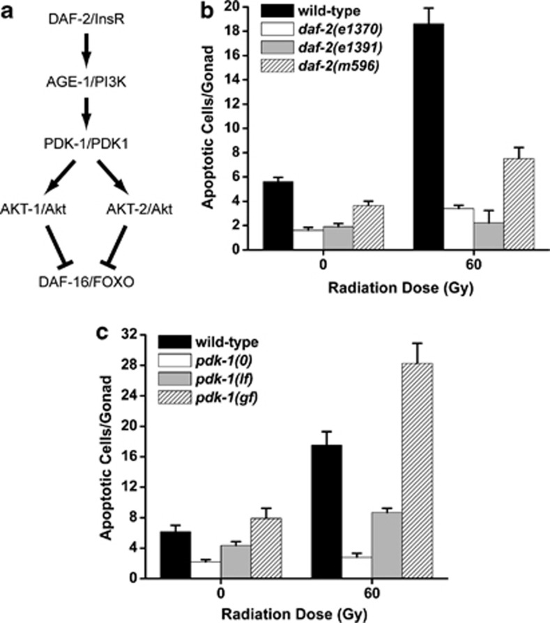
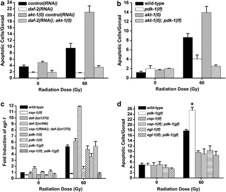
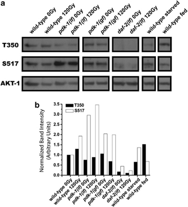
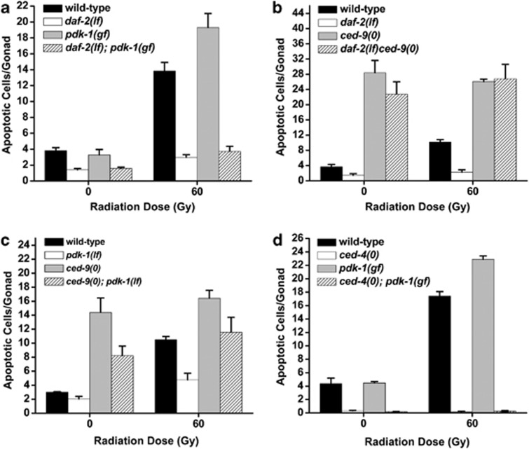
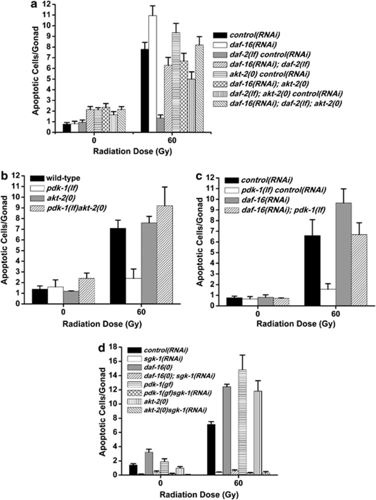
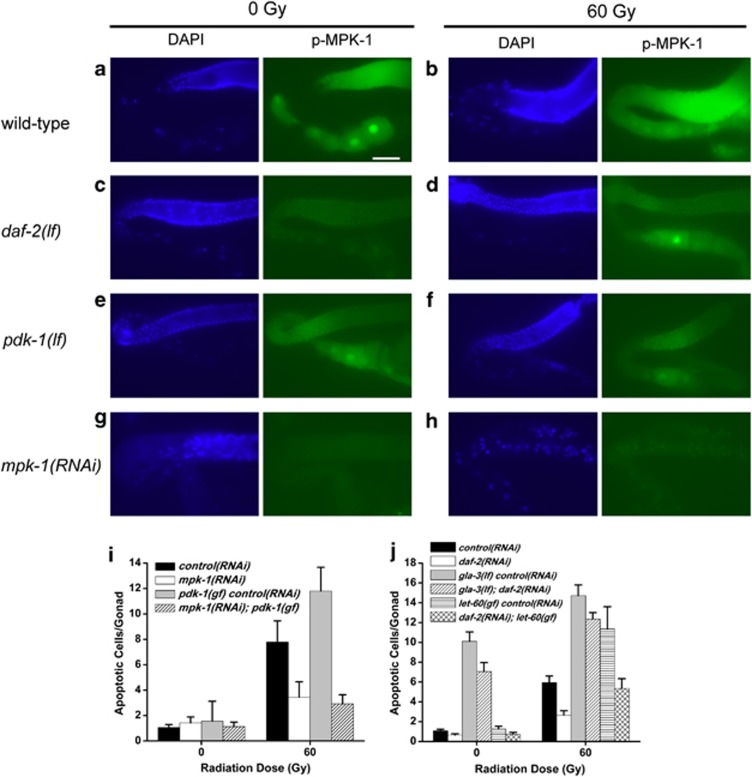
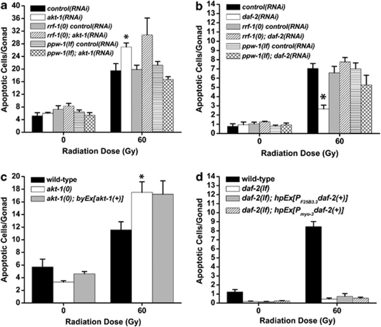
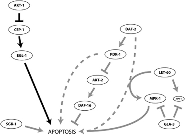
The insulin/IGF-1 pathway controls a number of physiological processes in the nematode worm Caenorhabditis elegans, including development, aging and stress response. We previously found that the Akt/PKB ortholog AKT-1 dampens the apoptotic response to genotoxic stress in the germline by negatively regulating the p53-like transcription factor CEP-1. Here, we report unexpected rearrangements to the insulin/IGF-1 pathway, whereby the insulin-like receptor DAF-2 and 3-phosphoinositide-dependent protein kinase PDK-1 oppose AKT-1 to promote DNA damage-induced apoptosis. While DNA damage does not affect phosphorylation at the PDK-1 site Thr350/Thr308 of AKT-1, it increased phosphorylation at Ser517/Ser473. Although ablation of daf-2 or pdk-1 completely suppressed akt-1-dependent apoptosis, the transcriptional activation of CEP-1 was unaffected, suggesting that daf-2 and pdk-1 act independently or downstream of cep-1 and akt-1. Ablation of the akt-1 paralog akt-2 or the downstream target of the insulin/IGF-1 pathway daf-16 (a FOXO transcription factor) restored sensitivity to damage-induced apoptosis in daf-2 and pdk-1 mutants. In addition, daf-2 and pdk-1 mutants have reduced levels of phospho-MPK-1/ERK in their germ cells, indicating that the insulin/IGF-1 pathway promotes Ras signaling in the germline. Ablation of the Ras effector gla-3, a negative regulator of mpk-1, restored sensitivity to apoptosis in daf-2 mutants, suggesting that gla-3 acts downstream of daf-2. In addition, the hypersensitivity of let-60/Ras gain-of-function mutants to damage-induced apoptosis was suppressed to wild-type levels by ablation of daf-2. Thus, insulin/IGF-1 signaling selectively engages AKT-2/DAF-16 to promote DNA damage-induced germ cell apoptosis downstream of CEP-1 through the Ras pathway.


Figures

Figure 1
Figure 1
PI3K signaling promotes DNA damage-induced germ cell apoptosis. (a) The PI3K pathway in C. elegans. (b and c) C. elegans PI3K signaling components, daf-2 and pdk-1, are required for damage-induced apoptosis. L4-stage worms were treated with IR and shifted to 25°C. Germ cell apoptosis was quantified 24 h later. pdk-1(0)=pdk-1(sa680), pdk-1(lf)=pdk-1(sa709), pdk-1(gf)=pdk-1(mg142). Error bars represent the S.E.M. from at least three independent experiments. At least ten animals were examined in each experiment
Figure 2
Figure 2
daf-2 and pdk-1 do not function upstream of akt-1 or cep-1. (a) Young adults were irradiated, incubated at 20°C for 24 h and apoptosis was quantified as in Figure 1. RNAi was performed in the rrf-3(pk1426) background. akt-1(0)=akt-1(ok525). (b) L4 stage worms were treated as in (a). pdk-1(lf)=pdk-1(sa709), akt-1(0)=akt-1(ok525). (c) L4 stage worms were treated with IR, incubated at 25°C for 24 h and total RNA was isolated. egl-1 transcript was quantified by Real-Time PCR using tbg-1 as an internal standard. cep-1(lf)=cep-1(gk138), pdk-1(0)=pdk-1(sa680), pdk-1(lf)=pdk-1(sa709), pdk-1(gf)=pdk-1(mg142). (d) L4 stage worms were treated with IR and germ cell apoptosis was quantified after 24 h at 25°C. pdk-1(gf)=pdk-1(mg142), cep-1(lf)=cep-1(gk138), egl-1(lf)=egl-1(n1084n3082). Error bars as in Figure 1. *P<0.001 versus wild-type
Figure 3
Figure 3
AKT-1 is regulated independently of Thr350 phosphorylation. (a) Worms were irradiated and AKT-1 was immunoprecipitated as described in Materials and Methods. All samples were run on the same gel, transferred to PVDF and probed for phospho-T350, S517 and total AKT-1. pdk-1(lf)=pdk-1(sa709); pdk-1(gf)=pdk-1(mg142); daf-2(lf)=daf-2(e1370). (b) The intensity of T350 and S517 in each lane was quantified using ImageJ and normalized to total AKT-1. The normalized intensity of each band was expressed relative to wild-type unirradiated. Data represent the average of two experiments
Figure 4
Figure 4
daf-2 and pdk-1 independently regulate the core apoptosis pathway. L4 stage (a, c, d) or young adult (b) worms were irradiated and germ cell apoptosis was quantified after 24 h at 20°C (b and c), or 25°C (a and d). Owing to the somatic effects of daf-2 and pdk-1 loss on the visibility of the germline in daf-2ced-9 and ced-9; pdk-1 double mutants, we found it necessary to perform these two epistasis experiments at slightly different developmental time points (48 h post-L4 in the case of daf-2ced-9 and 24 h post-L4 for ced-9; pdk-1). As worms age from L4 to young adult, the germline continues to expand in size and this is reflected by a small increase in basal levels of apoptosis. Therefore, the absolute levels of apoptosis seen in ced-9(0) mutants in the daf-2ced-9 experiment is greater than that seen in the ced-9; pdk-1 experiment. This should not alter our interpretation of epistasis, however, as loss of pdk-1 exerts an ∼1.3-fold greater suppressive effect on ced-9(0) than does loss of daf-2. daf-2(lf)=daf-2(e1370), ced-9(0)=ced-9(n2812), ced-4(0)=ced-4(n1162). Error bars as in Figure 1
Figure 5
Figure 5
daf-2 promotes damage-induced germ cell apoptosis via akt-2 and daf-16. (a–d) L4 stage worms were transferred to fresh RNAi plates, treated with IR and germ cell apoptosis quantified after 24 h at 20°C. akt-2(0)=akt-2(ok393), daf-2(lf)=daf-2(e1370), daf-16(0)=daf-16(mgDf47), pdk-1(lf)=pdk-1(sa709). Because the kinetics underlying apoptosis are increased at higher temperatures, the number of apoptotic cells observed in assays conducted at 20°C is lower than that seen at 25°C (see Figure 1). This reflects the temperature-dependent effects on the apoptotic process itself; the relative difference between genotypes is usually independent of this. Control RNAi experiments were carried out in wild-type (N2) animals
Figure 6
Figure 6
daf-2 and pdk-1 promote germline apoptosis through Ras/MAPK signaling. (a–f) Phospho-MPK-1 and DAPI staining in germlines of the indicated genotype before and after irradiation. (g–h) mpk-1(RNAi) negative controls in the absence and presence of IR. (i) Loss of mpk-1 in pdk-1(mg142) mutants suppresses excessive apoptosis. (j) Ablation of daf-2 by RNAi suppresses IR-induced germ cell apoptosis to wild-type levels in let-60(ga89) gf mutants, but does not suppress apoptosis in gla-3(op216) mutants. Worms were irradiated as in Figure 1 and incubated at 20°C for 24 h. Error bars as in Figure 1
Figure 7
Figure 7
akt-1 and daf-2 function from different tissues to regulate damage-induced germ cell apoptosis. (a) Young adult worms were transferred to fresh RNAi plates, treated with IR and germ cell apoptosis was quantified after 24 h at 20°C. rrf-1 and ppw-1 are required for RNAi in the soma and germline, respectively. rrf-1(0)=rrf-1(pk1417), ppw-1(lf)=ppw-1(pk1425). Error bars as in Figure 1. *P<0.05 versus control(RNAi). (b) L4-stage worms were transferred to fresh RNAi plates, treated with IR and germ cell apoptosis was quantified after 24 h at 20°C. Error bars as in Figure 1. *P<0.01 versus control(RNAi). (c) Young adult worms were treated with IR and germ cell apoptosis was quantified after 24 h at 20°C. byEx[akt-1(+)] is a high-copy extrachromosomal array that rescues the somatic Daf-c phenotype of akt-1(ok525); akt-2(RNAi) worms. akt-1(0)=akt-1(ok525). Error bars are as in Figure 1. *P<0.05 versus wild type. (d) L4-stage worms were transferred to fresh plates, treated with IR and germ cell apoptosis was quantified after 24 h at 20°C. daf-2(lf)=daf-2(e1370), hpEx[PF25B3.3daf-2(+)]=hpEx792, a high-copy array that expresses daf-2 in the nervous system; hpEx[Pmyo-3daf-2(+)]=hpEx791, a high-copy array that expresses daf-2 in muscle. Error bars are as in Figure 1
Figure 8
Figure 8
Model depicting the regulation of DNA damage-induced germ cell apoptosis by C. elegans PI3K pathway components. DAF-2 promotes apoptosis independently of AKT-1 by selectively engaging AKT-2 to regulate DAF-16 (AKT-1 inhibits CEP-1 from within damaged germ cells). PDK-1 functions through CED-4 and at least partially through AKT-2/DAF-16 to promote apoptosis downstream of CEP-1 and EGL-1. Although PDK-1 cannot promote apoptosis in the absence of CEP-1 and EGL-1, both DAF-2 and PDK-1 are required for amplification of LET-60/Ras signals that promote MPK-1/ERK phosphorylation and increase damage-induced apoptosis in the germline. SGK-1 promotes damage-induced germ cell apoptosis independently of the canonical Insulin/IGF-1 pathway. Arrows indicate activation, cross-hatches inhibition

References

    1. Vivanco I, Sawyers CL. The phosphatidylinositol 3-Kinase AKT pathway in human cancer. Nat Rev Cancer. 2002;2:489–501. - PubMed
    1. Liu P, Cheng H, Roberts TM, Zhao JJ. Targeting the phosphoinositide 3-kinase pathway in cancer. Nat Rev Drug Discov. 2009;8:627–644. - PMC - PubMed
    1. Contessa JN, Hampton J, Lammering G, Mikkelsen RB, Dent P, Valerie K, et al. Ionizing radiation activates Erb-B receptor dependent Akt and p70 S6 kinase signaling in carcinoma cells. Oncogene. 2002;21:4032–4041. - PubMed
    1. Brognard J, Clark AS, Ni Y, Dennis PA. Akt/protein kinase B is constitutively active in non-small cell lung cancer cells and promotes cellular survival and resistance to chemotherapy and radiation. Cancer Res. 2001;61:3986–3997. - PubMed
    1. Paradis S, Ailion M, Toker A, Thomas JH, Ruvkun G. A PDK1 homolog is necessary and sufficient to transduce AGE-1 PI3 kinase signals that regulate diapause in Caenorhabditis elegans. Genes Dev. 1999;13:1438–1452. - PMC - PubMed

Publication types

MeSH terms