. 2012 Oct 1;11(19):3568-77.
doi: 10.4161/cc.21869. Epub 2012 Aug 30.

Familial partial lipodystrophy, mandibuloacral dysplasia and restrictive dermopathy feature barrier-to-autointegration factor (BAF) nuclear redistribution

Affiliations

Familial partial lipodystrophy, mandibuloacral dysplasia and restrictive dermopathy feature barrier-to-autointegration factor (BAF) nuclear redistribution

Cristina Capanni et al. Cell Cycle. .

Abstract

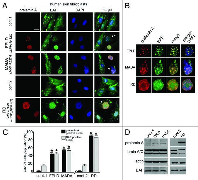
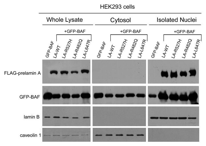
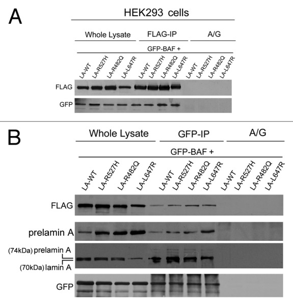
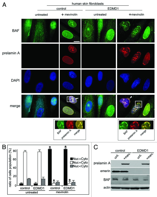
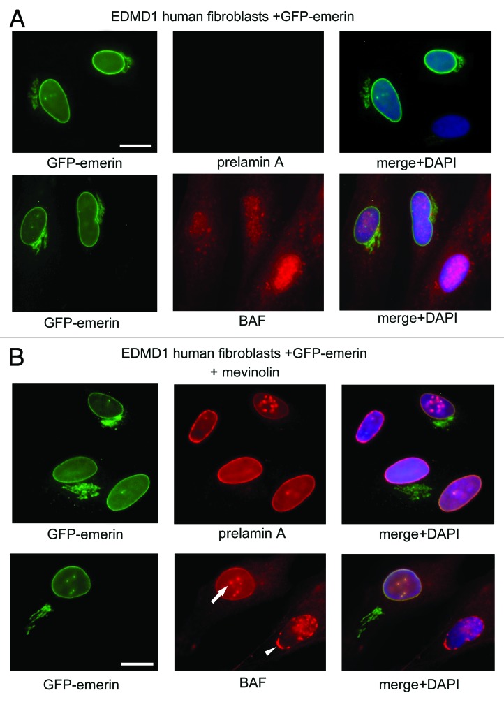
Prelamin A processing impairment is a common feature of a restricted group of rare genetic alterations/disorders associated with a wide range of clinical phenotypes. Changes in histone posttranslational modifications, alterations in non-histone chromatin proteins and chromatin disorganization have been specifically linked to impairment of specific, distinct prelamin A processing steps, but the molecular mechanism involved in these processes is not yet understood . In this study, we show that the accumulation of wild-type prelamin A detected in restrictive dermopathy (RD), as well as the accumulation of mutated forms of prelamin A identified in familial partial lipodystrophy (FPLD) and mandibuloacral dysplasia (MADA), affect the nuclear localization of barrier-to-autointegration factor (BAF), a protein able to link lamin A precursor to chromatin remodeling functions. Our findings, in accordance with previously described results, support the hypothesis of a prelamin A involvement in BAF nuclear recruitment and suggest BAF-prelamin A complex as a protein platform usually activated in prelamin A-accumulating diseases. Finally, we demonstrate the involvement of the inner nuclear membrane protein emerin in the proper localization of BAF-prelamin A complex.


Figures

None
Figure 1. Barrier-to-autointegration factor nuclear localization in prelamin A-accumulating diseases. (A) BAF and prelamin A distribution in control (cont.1 adult healthy donor, cont.2 neonatal healthy donor) Familial partial lipodystrophy (FPLD, LMNA-R482Q), Mandibuloacral dysplasia (MADA, LMNA-R527H) and Restrictive dermopathy (RD, ZMPSTE24 c.1085_1086InsT). Control and pathological cells at early passage (passage 5–10) were used for each experiment. BAF was evaluated on methanol-fixed cells by a rabbit-polyclonal anti-BAF antibody visualized by FITC-conjugated secondary antibody (green). Prelamin A was evaluated using a goat-polyclonal anti-prelamin A antibody visualized by TRITC-conjugated secondary antibody (red). In merge, arrow, arrowhead and asterisk indicate double-stained nuclei. DNA was detected using DAPI. Bar, 10 μm. (B) Higher magnification of prelamin A-BAF-positive nuclei indicated by arrow, arrowhead and asterisk in panel A. Prelamin A (red), BAF (green) merge (merge) and merge plus DAPI staining (merge + DAPI) are shown. (C) The percentage of nuclei showing prelamin A staining is reported in the graph as means ± SD of three different counts (100 nuclei per count). Examined samples are control fibroblast from adult healthy donors (cont.1), familial partial lipodystrophy fibroblasts (FPLD), mandibuloacral dysplasia fibroblasts (MADA), control fibroblast from neonate healthy donors (cont.2) and restrictive dermopathy fibroblasts (RD). Asterisks indicate statistically significant differences at the Student’s t-test (p < 0.05), with respect to control cells values. (D) Western blotting evaluation of BAF protein levels in control (cont.1 and cont.2), FPLD, MADA and RD cells. Eighty micrograms of total cell lysates were subjected to western blot detection of prelamin A (prelamin A), lamin A/C (lamin A/C), BAF (BAF) and actin (actin). Immunolabeled bands visualized by ECL chemiluminescence are shown.
None
Figure 2. Barrier-to-autointegration factor localization and expression in prelamin A mutants HEK293 transfected cells. (A) BAF localization in HEK293 cells expressing prelamin A mutants. Cells were transfected with wild-type processable prelamin A (LA-WT), MADA processable prelamin A mutant (LA-R527H), FPLD processable prelamin A mutant (LA-R482Q) or uncleavable farnesylated-carboxymethylated prelamin A mutant (LA-L647R). Immunofluorescence detection of overexpressed proteins was performed using a mouse monoclonal anti FLAG-Cy3 conjugated antibody (red). Arrowhead and arrow indicate LA-R527H and LA-R482Q distribution, respectively. Endogenous BAF localization was evaluated by a rabbit polyclonal anti-BAF antibody visualized by FITC-conjugated secondary antibody (green). Asterisk indicates BAF distribution in untransfected cells. Nuclei were stained with DAPI. Bar, 10 μm. (B) BAF protein levels in prelamin A mutants expressing cells. BAF protein levels were determined in HEK293 cells expressing FLAG-tagged prelamin A constructs (LA-WT, LA-R527H, LA-R482Q, LA-L647R) as well as in untransfected cells (unt.). Thirty micrograms of total cell lysates were separated by SDS-PAGE (5–20%) and subjected to western blot. FLAG-tagged protein and endogenous BAF detection was performed using a mouse-monoclonal and a rabbit-polyclonal, respectively. Actin (Actin) was detected as a protein-loading control.
None
Figure 3. Barrier-to-autointegration factor nuclear translocation in HEK293 cells transfected with prelamin A mutants. Western blotting analysis of whole cellular lysates, cytosol and isolated nuclei from HeK293 cells transfected with GFP-BAF (GFP-BAF), alone or in combination with: wild-type processable prelamin A (LA-WT + GFP-BAF), MADA processable prelamin A mutant (LA-R527H+GFP-BAF), FPLD processable prelamin A mutant (LA-R482Q+GFP-BAF) or uncleavable prelamin A mutant (LA-L647R+GFP-BAF). Immunolabeled bands of FLAG, GFP, lamin B1 and caveolin 1 are shown.
None
Figure 4. Barrier-to-autointegration factor interaction with laminopathic forms of prelamin A. HEK293 cells were transfected with FLAG-tagged prelamin A constructs (LA-WT, LA-R527H, LA-R482Q, LA-L647R) in combination with GFP-BAF (GFP-BAF) construct. Cotransfected cells were lysed and subjected to coimmunoprecipitation experiments. Total lysates (whole lysate) and immunoprecipitated proteins, FLAG-IP in panel (A) and GFP-IP in panel (B), were subjected to western blotting analysis. FLAG and GFP immunolabeled bands were observed in whole lysates as well as in immunoprecipitated samples, while no protein staining was observed in coimmunoprecipitation samples obtained in absence of anti-FLAG antibody or anti GFP- antibody (A/G) (A and B). In (B), prelamin A staining was observed in whole lysates and in GFP-IP lanes subjected to specific anti-prelamin A detection (prelamin A) and to anti-lamin A detection performed with Abcam antibody, which detects both mature lamin A (70kDa) and prelamin A (74kDa) (prelamin A, lamin A).
None
Figure 5. Localization and expression of barrier-to-autointegration factor and prelamin A in EDMD1 cells. (A) BAF and prelamin A were localized in control cells (control) treated with 10 μM mevinolin (+mevinolin) and in Emery Dreifuss Muscular Dystrophy type 1 (EDMD1) cells untreated (untreated) or treated (+mevinolin) with 10 μM mevinolin. After pharmacological treatment, BAF (green) and prelamin A staining (red) were performed. BAF and prelamin A were detected using a rabbit-polyclonal antibody and a goat-polyclonal antibody, respectively. Prelamin A and BAF colocalization is indicated by arrow and arrowhead in merge (merge). Squares indicate BAF/prelamin A aggregates localization. In the lower panels, BAF/prelamin A aggregates localization is reported at higher magnification, omitting DAPI staining. DNA was stained by DAPI. Bar, 10 μm. (B) Proportions of cell populations with different BAF localization: Nuc > Cyto, Nuc = Cyto and Nuc < Cyto represent cells with prevailing nuclear staining, uniform or mostly cytoplasmic staining, respectively. The proportion of cell populations was determined in control and EDMD1 cells at the same population doubling untreated (untreated) or mevinolin-treated cells (mevinolin). Data are means ± s.d. of at least 100 cells for each of three independent experiments. Asterisks indicate statistically significant differences at the Student’s t-test (p < 0.05), with respect to control and EDMD1 mevinolin untreated cells values. (C) Western blotting evaluation of BAF in prelamin A EDMD1-accumulating cells. Western blotting evaluation of prelamin A, emerin, BAF and actin were performed in total lysates from control cells (control) or Emery Dreifuss Muscular Dystrophy type 1 cells (EDMD1) untreated (unt.) or treated with mevinolin (+mev.). Immunolabeled bands are shown.
None
Figure 6. Emerin exogenous expression rescues BAF intranuclear distribution in EDMD1 cells accumulating prelamin A. Prelamin A and BAF evaluation in EDMD1 GFP-emerin transfected cells with or without mevinolin treatment. In panel A, untreated EDMD1 GFP-emerin transfected cells (green) were subjected to prelamin A (prelamin A) and BAF (BAF) detection, both protein were visualized by TRITC-conjugated secondary antibody (red). In merge (merge+DAPI) DNA was detected using DAPI. Bar, 10 μm. In panel B, mevinolin-treated EDMD1 GFP-emerin transfected cells (green). Prelamin A (prelamin A) and BAF (BAF) localization are shown. Arrow indicates recover of intranuclear BAF distribution in GFP-emerin transfected cells. Arrowhead indicates the altered intranuclear BAF distribution in the absence of emerin.

Similar articles

Cited by

References

    1. Misteli T. Higher-order genome organization in human disease. Cold Spring Harb Perspect Biol. 2010;2:a000794. doi: 10.1101/cshperspect.a000794. - DOI - PMC - PubMed
    1. Worman HJ. Nuclear lamins and laminopathies. J Pathol. 2012;226:316–25. doi: 10.1002/path.2999. - DOI - PMC - PubMed
    1. Maraldi NM, Lattanzi G, Capanni C, Columbaro M, Merlini L, Mattioli E, et al. Nuclear envelope proteins and chromatin arrangement: a pathogenic mechanism for laminopathies. Eur J Histochem. 2006;50:1–8. - PubMed
    1. Maraldi NM, Lattanzi G. Involvement of prelamin A in laminopathies. Crit Rev Eukaryot Gene Expr. 2007;17:317–34. doi: 10.1615/CritRevEukarGeneExpr.v17.i4.50. - DOI - PubMed
    1. Sinensky M, Fantle K, Trujillo M, McLain T, Kupfer A, Dalton M. The processing pathway of prelamin A. J Cell Sci. 1994;107:61–7. - PubMed

Publication types

MeSH terms

Supplementary concepts

LinkOut - more resources