

doi: 10.4161/cc.21927.
Epub 2012 Aug 30.
VDAC3 regulates centriole assembly by targeting Mps1 to centrosomes
Affiliations
- PMID: 22935710
- PMCID: PMC3478317
- DOI: 10.4161/cc.21927
Item in Clipboard
VDAC3 regulates centriole assembly by targeting Mps1 to centrosomes
Cell Cycle.
.
Abstract
Centrioles are duplicated during S-phase to generate the two centrosomes that serve as mitotic spindle poles during mitosis. The centrosomal pool of the Mps1 kinase is important for centriole assembly, but how Mps1 is delivered to centrosomes is unknown. Here we have identified a centrosome localization domain within Mps1 and identified the mitochondrial porin VDAC3 as a protein that binds to this region of Mps1. Moreover, we show that VDAC3 is present at the mother centriole and modulates centriole assembly by recruiting Mps1 to centrosomes.
Figures
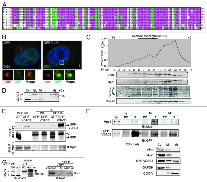
Figure 1. VDAC3 physically interacts with Mps1. (A) Conservation in the Mps1 N terminus; highlighted residues are identical (pink) or conserved in structure or function (green) in at least 75% of the species. H.s., Homo sapiens (human); B.s., Bos taurus (bovine); C.f., Canis familiaris (dog); G.g., Gallus gallus (chicken); O.c., Oryctolagus cuniculus (European rabbit); M.m., Mus musculus (mouse); X.l., Xenopus laevis (frog); D.r., Danio rerio (zebrafish). (B) S-phase arrested HeLa cells expressing GFP or GFP-Mps153-175 (GFP-CLD) were stained for γ-tubulin (γ-tub; red) and DNA (blue). Bar = 5 μm. In this and other figures, panels show 4-fold digitally magnified images of a region of interest, in this case surrounding the centrosomes. (C) Sucrose gradient fractionation of nucleui-depleted extract from HeLa cells, where the graph represents the total protein concentrations of the fractions and the immunoblots show the distribution of indicated protein in these fractions. The absence of nuclear protein contamination was verified by immunoblotting with anti-LaminB antibody (data not shown). (D) Differential distribution of VDAC3a and VDAC3b as shown by immunoblotting of subcellular fractions [cytosolic (Cy), mitochondrial (Mt), microsomal (Mi) and pellet (N/u)] obtained using Qproteome mitochondria isolation protocol. (E) After pre-clearing with beads alone (PC), anti-GFP immunoprecipitates from S-phase arrested HEK293 cells expressing GFP or GFP-VDAC3 (arrowheads) were immunoblotted with antibodies against Mps1 and GFP. Caret indicates Protein G. (F) After pre-clearing (PC), GFP-VDAC3 immunoprecipitates (IP) from the subcellular fractions from S-phase arrested HEK293 cells expressing GFP-VDAC3 were immunoblotted as in (D). Either 1% (cytosol) or 2% (other fractions) of the input (In) were analyzed with the indicated antibodies. Although a truncated form of GFP-VDAC3 was detected in lysates of cells expressing GFP-VDAC3 (see Fig. S1F), only the band of expected size (full-length GFP-VDAC3) was shown for convenience, in (E and F). (G) After pre-clearing (PC), anti-Mps1 immunoprecipitates from S-phase arrested RPE1 cells were separated in reducing (+DTT) or non-reducing (-DTT) condition and immunoblotted with indicated antibodies. Trace amount of Protein G and IgG light chain are indicated by caret and arrowhead, respectively.
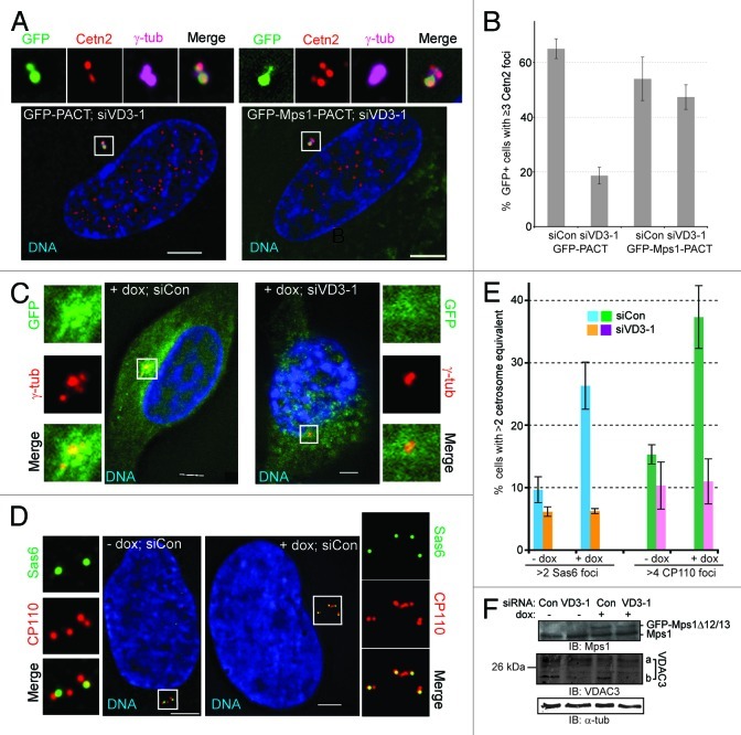
Figure 5. VDAC3 recruits Mps1 to centrosomes to regulate centriole assembly. (A and B) siCon or siVDAC3–1 (siVD3–1) RPE1 cells expressing GFP-PACT (green) or GFP-Mps1-PACT (green) were arrested in S-phase by a 24 h HU treatment and were examined for centriole numbers. (A) Shown are representative siVDAC3–1 cells stained for Cetn2 (red), γ-tub (magenta) and DNA (blue); GFP is green, bar = 5 μm. (B) Percentage of GFP-positive cells with ≥ 3 Cetn2 foci, values represent the mean ± SD for three independent experiments, 50–60 cells counted per replicate. (C–F) HeLa GFP-Mps1∆12/13 cells transfected with control (siCon) and VDAC3–1 (siVD3–1) siRNA were arrested in S-phase by a 72 h HU treatment, with or without dox, to induce GFP-Mps1∆12/13 [green in (C)], and stained for DNA (blue) and (C) γ-tub (red) or (D) CP110 (red) and Sas6 (green). Bar = 5 μm. (E) The percentage of cells with more than two centrosome equivalents (> 2 Sas6 foci or > 4 CP110 foci), values represent the mean ± SD for three independent experiments, at least 100 cells counted per replicate. (F) Immunoblots showing the expression of GFP-Mps1∆12/13 (arrowhead), endogenous Mps1 (arrow) and depletion of VDAC3 (VDAC3a roughly 10%, VDAC3b roughly 80%), α-tub as loading control.

Figure 3. VDAC3 recruits Mps1 to centrosomes. (A and B) Representative RPE1 cells stained for Mps1 (green) and (A) Ninein (red) or (B) VDAC3 (red). DNA is blue, bar = 5 μm. (C and D) Asynchronously growing RPE1 cells transfected with control (Con) or VDAC3 siRNA-1 (VD3–1) analyzed by (C) qRT-PCR or (D) immunoblot. (C) Relative expression of VDAC3 mRNA, values represent mean ± SD for three replicates. (D) Immunoblot showing VDAC3a decreased by roughly 35%, and VDAC3b by 75%. GAPDH was used as loading control. (E and F) Representative images of RPE1 cells prepared as in (C and D) stained for VDAC3 (red) and (E) Ac-tub (green) or (F) Cep170 (green). DNA is blue, bar = 5 μm. Panels show magnified centrosomes/basal bodies. In (F), cartoon shows localization of VDAC3 (red) with respect to Cep170 (green) in control and VDAC3-depleted RPE1 cells. (G) Micrographs show representative images of RPE1 cells prepared as in (C and D), treated with DMSO or MG115, labeled with BrdU for 4 h and stained for Mps1 (green), γ-tub (red) and BrdU (blue). Panels show magnified centrosomes. Bar = 5 μm. Immunoblots show whole-cell level of Mps1, α-tubulin (α-tub) as loading control. Centrosomal Mps1 level was determined as described in Materials and Methods. Values represent mean ± SD of 25 representative cells.
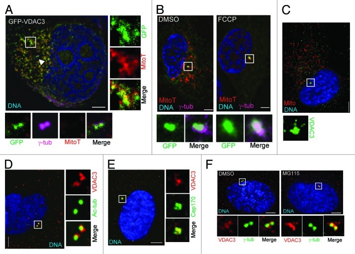
Figure 2. VDAC3 is a centrosomal protein of the mother centriole. (A) A representative HeLa cell expressing GFP-VDAC3 (green) stained with MitotrackerRed (MitoT, red), then fixed and stained for γ-tub (magenta). Arrowhead indicates centrosomes, box indicates a region containing mitochondria. (B) RPE1 cells expressing GFP-VDAC3 (green) were treated with either DMSO (solvent control) or 200 μM FCCP for 1 h and stained with MitoT (red). Shown are representative images stained for γ-tub (magenta). (C–E) Asynchronously growing RPE1 cells were stained for (C) VDAC3 (green) and mitochondria (Mito, red), (D) VDAC3 (red) and Ac-tubulin (green), and (E) VDAC3 (red) and Cep170 (green). Shown are representative images where the predominant centrosomal VDAC3 signal was at both centrosomes (C and D) or at the mother centriole (E). (F) Representative images of asynchronously growing RPE1 cells treated with either DMSO or MG115 for 4 h, stained for γ-tub (green), VDAC3 (red) and imaged under identical condition. DNA is blue and bar is 5 μm in (A–F).
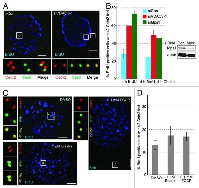
Figure 4. Depletion of VDAC3 or Mps1 inhibits centriole assembly. (A and B) Asynchronously growing RPE1 cells transfected with control (siCon), VDAC3- (siVDAC3–1) or Mps1-specific (siMps1) siRNAs were labeled with a 4 h pulse of BrdU. Centriole number was determined after this pulse (4 h BrdU), or after an additional 4 h chase (4 h BrdU, 4 h chase) using antibodies against Cetn2 and Sas6. (A) Representative images of siCon or siVDAC3–1 cells stained for Cetn2 (red), Sas6 (green) and BrdU (blue). Bar = 5 μm. (B) Percentage of BrdU-positive cells with ≤ 2 Cetn2 foci. Values represent mean ± SD for three independent experiments, 75–100 cells counted per replicate. Immunoblots show depletion of Mps1 (by roughly 85%), α-tub as loading control. (C and D) Growing RPE1 cells were treated with DMSO, 1 μM Erastin for 16 h or 100 μM FCCP for 4 h, labeled with a 4 h pulse of BrdU and stained for Cetn2. (C) Shown are BrdU (blue), Cetn2 (red) and γ-tub (green) for representative cells. Bar = 5 μm. (D) Percentage of BrdU-positive cells with ≤ 2 Cetn2 foci, values represent mean ± SD for three independent experiments, 75–100 cells counted per replicate.
Similar articles
-
VDAC3 and Mps1 negatively regulate ciliogenesis.Cell Cycle. 2013 Mar 1;12(5):849-58. doi: 10.4161/cc.23824. Epub 2013 Feb 6. Cell Cycle. 2013. PMID: 23388454 Free PMC article.
-
Mps1 phosphorylation sites regulate the function of centrin 2 in centriole assembly.Mol Biol Cell. 2010 Dec;21(24):4361-72. doi: 10.1091/mbc.E10-04-0298. Epub 2010 Oct 27. Mol Biol Cell. 2010. PMID: 20980622 Free PMC article.
-
Antizyme restrains centrosome amplification by regulating the accumulation of Mps1 at centrosomes.Mol Biol Cell. 2010 Nov 15;21(22):3878-89. doi: 10.1091/mbc.E10-04-0281. Epub 2010 Sep 22. Mol Biol Cell. 2010. PMID: 20861309 Free PMC article.
-
Leader of the SAC: molecular mechanisms of Mps1/TTK regulation in mitosis.Open Biol. 2018 Aug;8(8):180109. doi: 10.1098/rsob.180109. Open Biol. 2018. PMID: 30111590 Free PMC article. Review.
-
The PLK4-STIL-SAS-6 module at the core of centriole duplication.Biochem Soc Trans. 2016 Oct 15;44(5):1253-1263. doi: 10.1042/BST20160116. Biochem Soc Trans. 2016. PMID: 27911707 Free PMC article. Review.
Cited by
-
Quantitative immunofluorescence assay to measure the variation in protein levels at centrosomes.J Vis Exp. 2014 Dec 20;(94):52030. doi: 10.3791/52030. J Vis Exp. 2014. PMID: 25548932 Free PMC article.
-
Identification of a new aggressive axis driven by ciliogenesis and absence of VDAC1-ΔC in clear cell Renal Cell Carcinoma patients.Theranostics. 2020 Feb 3;10(6):2696-2713. doi: 10.7150/thno.41001. eCollection 2020. Theranostics. 2020. PMID: 32194829 Free PMC article.
-
Modular elements of the TPR domain in the Mps1 N terminus differentially target Mps1 to the centrosome and kinetochore.Proc Natl Acad Sci U S A. 2016 Jul 12;113(28):7828-33. doi: 10.1073/pnas.1607421113. Epub 2016 Jun 23. Proc Natl Acad Sci U S A. 2016. PMID: 27339139 Free PMC article.
-
Centrin 3 is an inhibitor of centrosomal Mps1 and antagonizes centrin 2 function.Mol Biol Cell. 2015 Nov 1;26(21):3741-53. doi: 10.1091/mbc.E14-07-1248. Epub 2015 Sep 9. Mol Biol Cell. 2015. PMID: 26354417 Free PMC article.
-
MPS1 is involved in the HPV16-E7-mediated centrosomes amplification.Cell Div. 2021 Nov 4;16(1):6. doi: 10.1186/s13008-021-00074-9. Cell Div. 2021. PMID: 34736484 Free PMC article.
References
Publication types
MeSH terms
Substances
Grants and funding
LinkOut - more resources
Full Text Sources
Other Literature Sources
Miscellaneous