

Review
doi: 10.1102/1470-7330.2012.0012.
Advanced magnetic resonance imaging biomarkers of cerebral metastases
Affiliations
- PMID: 22935843
- PMCID: PMC3458786
- DOI: 10.1102/1470-7330.2012.0012
Item in Clipboard
Review
Advanced magnetic resonance imaging biomarkers of cerebral metastases
Cancer Imaging.
.
Abstract
There are a number of magnetic resonance imaging techniques available for use in the diagnosis and management of patients with cerebral metastases. This article reviews these techniques, in particular, the advanced imaging methodologies from which quantitative parameters can be derived, the role of these imaging biomarkers have in distinguishing metastases from primary central nervous system tumours and tumour mimics, and metrics that may be of value in predicting the origin of the primary tumour.
Figures
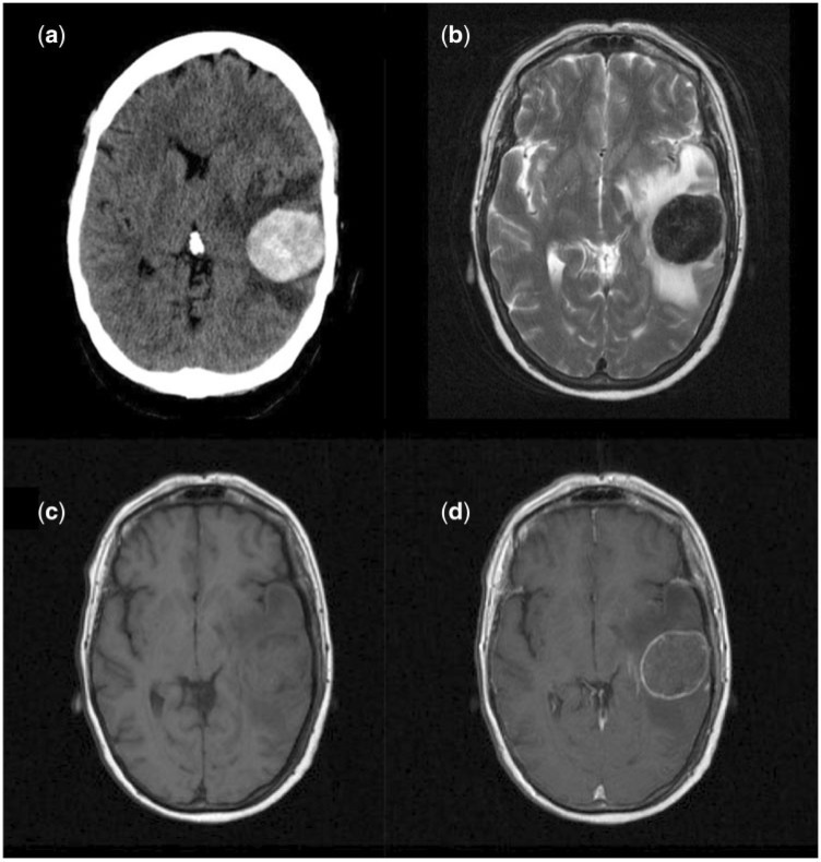
Imaging appearances of a solitary mucinous metastases from a colonic carcinoma primary on (a) non-contrast CT (hyper-attenuating lesion); (b) T2-weighted imaging (markedly hypointense); (c) T1-weighted pre-contrast imaging (isointense); (d) post-gadolinium contrast T1-weighted imaging (rim enhancement).
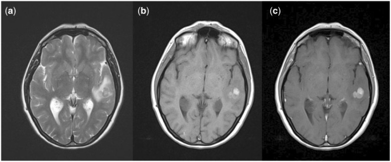
Imaging characteristics of melanoma metastases. (a) T2; (b) pre-contrast T1; (c) post-contrast T1.
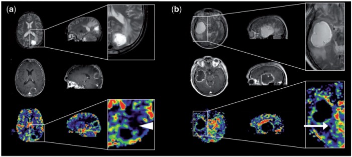
Perfusion imaging in (a) solitary cerebral metastasis and (b) GBM (axial imaging on the left and sagittal reformats on the right). Top row: T2-weighted imaging. Middle row: post-contrast T1-weighted imaging. Bottom row: CBV maps. Magnified images of CBV maps and corresponding T2-weighted images. (a) The peri-lesional non-enhancing tissue surrounding the metastasis exhibits a very low relative CBV (white arrowhead) and represents vasogenic oedema; (b) the peri-lesional non-enhancing tissue surrounding the GBM has a slightly higher relative cerebral blood volume (white arrow) and represents infiltrating glioma.
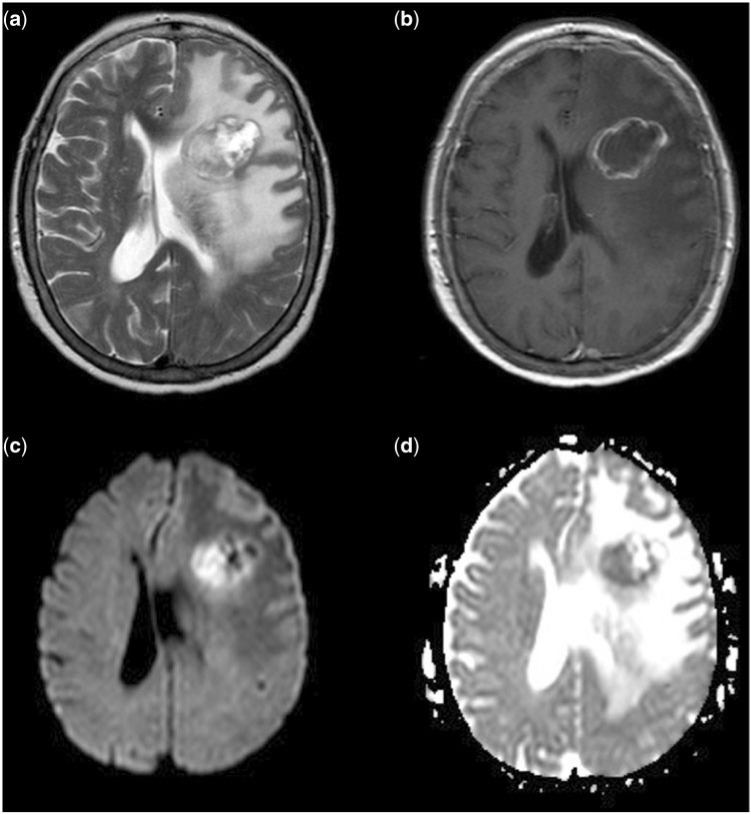
Restricted diffusion in cerebral metastases. (a) T2-weighted imaging; (b) post-contrast T1-weighted imaging; (c) DWI; (d) ADC map. There is restricted diffusion within the medial aspect of the tumour which demonstrates restriction in the more solid, T2 hypointense, non-enhancing component.
References
-
- Posner JB, Chernik NL. Intracranial metastases from systemic cancer. Adv Neurol. 1978;19:579–92. - PubMed
Publication types
MeSH terms
Substances
LinkOut - more resources
Full Text Sources
Medical