. 2012 Nov 1;186(9):866-76.
doi: 10.1164/rccm.201204-0754OC. Epub 2012 Aug 30.

Acellular normal and fibrotic human lung matrices as a culture system for in vitro investigation

Affiliations

Acellular normal and fibrotic human lung matrices as a culture system for in vitro investigation

Adam J Booth et al. Am J Respir Crit Care Med. .

Abstract

Rationale: Extracellular matrix (ECM) is a dynamic tissue that contributes to organ integrity and function, and its regulation of cell phenotype is a major aspect of cell biology. However, standard in vitro culture approaches are of unclear physiologic relevance because they do not mimic the compositional, architectural, or distensible nature of a living organ. In the lung, fibroblasts exist in ECM-rich interstitial spaces and are key effectors of lung fibrogenesis.

Objectives: To better address how ECM influences fibroblast phenotype in a disease-specific manner, we developed a culture system using acellular human normal and fibrotic lungs.

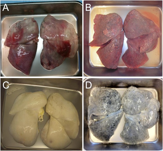
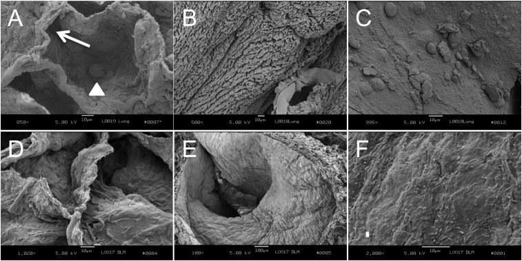
Methods: Decellularization was achieved using treatment with detergents, salts, and DNase. The resultant matrices can be sectioned as uniform slices within which cells were cultured.

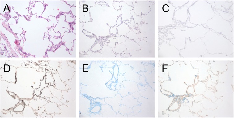
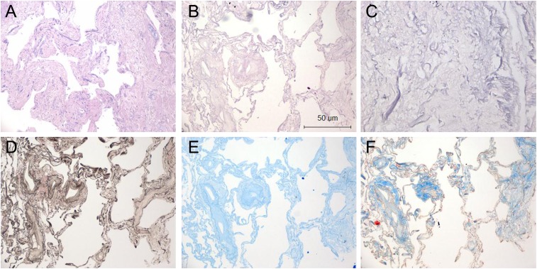
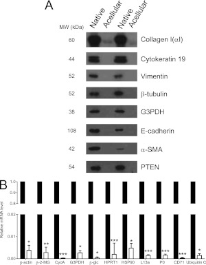
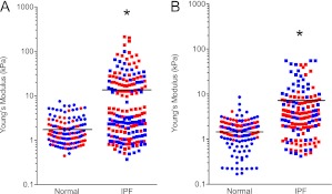
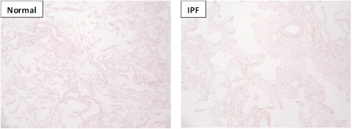
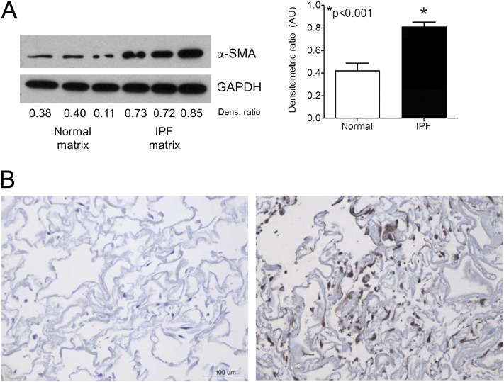
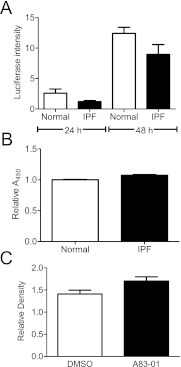
Measurements and main results: We report that the decellularization process effectively removes cellular and nuclear material while retaining native dimensionality and stiffness of lung tissue. We demonstrate that lung fibroblasts reseeded into acellular lung matrices can be subsequently assayed using conventional protocols; in this manner we show that fibrotic matrices clearly promote transforming growth factor-β-independent myofibroblast differentiation compared with normal matrices. Furthermore, comprehensive analysis of acellular matrix ECM details significant compositional differences between normal and fibrotic lungs, paving the way for further study of novel hypotheses.

Conclusions: This methodology is expected to allow investigation of important ECM-based hypotheses in human tissues and permits future scientific exploration in an organ- and disease-specific manner.


Figures

Figure 1.
Figure 1.
Histologic evaluation of native and acellular human normal lungs. (A) Native human normal lung, hematoxylin and eosin (H&E) stain. (B and C) Decellularized human lung stained with H&E at 10× (B) and 40× (C) magnification. (D) Verhoeff elastin stain, (E) Alcian blue, and (F) Masson trichrome stain (all 10× magnification). The decellularization process leaves the lung architecture intact.
Figure 2.
Figure 2.
Histologic evaluation of native and acellular human idiopathic pulmonary fibrosis (IPF) lungs. (A) Native human IPF lung, hematoxylin and eosin (H&E) stain. (B and C) Decellularized human IPF lung stained with H&E at 10× (B) and 40× (C) magnification. (D) Verhoeff elastin stain, (E) Alcian blue, and (F) Masson trichrome stain (all 10× magnification). The decellularization process leaves the lung architecture intact.
Figure 3.
Figure 3.
Confirmation of cellular protein and DNA removal. (A) Western blot analysis for cellular proteins indicated in right margin before (lanes 1 and 3) and after (lanes 2 and 4) decellularization; n = 2 separate lung samples. Results are representative of three Western blots. (B) Real-time semiquantitative polymerase chain reaction of various housekeeping genes in non-decellularized lung (closed bars) and decellularized lung matrix (open bars). Relative expression of each gene was less than 99.95% in decellularized matrix compared with native lung. Results are pooled from two separate experiments. α-SMA = α-smooth muscle actin; β-2-MG = β-2-microglobulin; β-glc = β-glucuronidase; CD71 = transferrin receptor; CycA = cyclophilin A; G3PDH = glyceraldehyde-3-phosphate dehydrogenase; HPRT1 = hypoxanthine phosphoribosyltransferase 1; HSP90 = heat shock protein 90 kD α (cytosolic); L13a = ribosomal protein L13a; P0 = ribosomal protein, large; PTEN = phosphatase and tensin homolog deleted on chromosome 10. *P < 0.005, **P < 0.01, ***P < 0.001.
Figure 4.
Figure 4.
Gross appearance of human lungs before and after decellularization. Normal lungs deemed unsuitable for transplantation and idiopathic pulmonary fibrosis lungs before (A and B, respectively) and after (C and D, respectively) decellularization. Lungs retain their normal gross structure and anatomic relationships.
Figure 5.
Figure 5.
Scanning electron microscopy of normal lung matrices before (AC) and after (DF) decellularization. (A) Normal alveolus. White arrow shows normal-appearing alveolar wall. Arrowhead shows an alveolar macrophage; 850× magnification. (B) Ciliated bronchus; 500× magnification. (C) Pulmonary blood vessel. Blood cells are adherent to the wall; 995× magnification. (D) Acellular alveoli; 1,020× magnification. (E) Acellular bronchus without ciliated lining cells; 100× magnification. (F) Acellular vessel without lining cells; 2,000× magnification. Scale bar = 10 μm.
Figure 6.
Figure 6.
Transmission electron microscopy of normal and idiopathic pulmonary fibrosis (IPF) acellular lung matrices. In normal matrix (left panel), alveolar epithelial and vascular endothelial basement membranes (black arrows) are of uniform thickness, and the interstitial space has organized fibers of collagen (white arrows) and discrete bundles of elastin (white arrowheads). In contrast, IPF matrix (right panel) shows a markedly thickened, heterogeneous basement membrane (black arrows) and haphazardly located, disorganized collagen fibers (white arrows) with disorganized elastin fragments (white arrowhead). No cellular material is observed. A = alveolar space. V = vascular space. Both panels, 8,520× magnification. Scale bar = 2 μm.
Figure 7.
Figure 7.
Stiffness of native and acellular normal and idiopathic pulmonary fibrosis (IPF) lung matrices as determined by atomic force microscopy (n = 2 samples per condition; each color represents a different individual). (A) Native normal human lung possessed a mean (± SEM) Young’s modulus of 1.96 ± 0.13 kPa, whereas native IPF lung possessed a mean (± SEM) stiffness of 16.52 ± 2.25 kPa, significantly higher than normal tissue (P < 0.0001 by Mann-Whitney test). (B) The mean (± SEM) elastic (Young’s) modulus of decellularized normal lung is approximately 1.6 (± 0.08) kPa. By comparison, the mean (± SEM) Young’s modulus of decellularized IPF lung was 7.34 ± 0.6 kPa. Each point represents a single measurement of a 30 × 30 μm2 area. *P < 0.0001.
Figure 8.
Figure 8.
Hematoxylin and eosin stain (10× magnification) of normal and idiopathic pulmonary fibrosis lung tissue immediately adjacent to tissue assessed by liquid chromatography–tandem mass spectrometry reported in Table 1.
Figure 9.
Figure 9.
Idiopathic pulmonary fibrosis (IPF) matrices induce α-smooth muscle actin (α-SMA) expression in normal human lung fibroblasts. (A) Western blot analysis of normal human lung fibroblasts cultured within human acellular normal or IPF lung matrix. The same cells at the same passage were cultured in matrices for 48 hours before harvesting. IPF lung matrix significantly induced α-SMA. Bar graphs depict the mean (± SEM) densitometric ratio of α-SMA normalized to glyceraldehyde-3-phosphate dehydrogenase (GAPDH). The blot is representative of triplicate experiments using three different primary cell lines and matrices derived from three separate individuals. (B) α-SMA immunohistochemistry of normal fibroblasts seeded within normal (left panel) or IPF (right panel) matrices for 48 hours; 20× magnification.
Figure 10.
Figure 10.
Enhanced α-smooth muscle actin expression in fibroblasts in idiopathic pulmonary fibrosis (IPF) matrices is transforming growth factor (TGF)-β–independent. (A) Normal human lung fibroblasts were cultured in normal or IPF matrices for 24 hours in the upper well of a transwell system, and 1 × 10⁵ mink lung epithelial cells (MLECs) were cultured in the lower chamber. Lysates of MLECs were assayed by a luminometer and relative intensity was plotted above. There was no difference in TGF-β activity released from normal or IPF matrices. Results are pooled from three separate experiments each performed in triplicate tissue slices. (B) Intact acellular matrices were acid activated and the resultant media was assayed for TGF-β levels by enzyme immunoassay. No difference in total TGF-β was observed between normal and IPF slices. Data are the pooled mean (± SD) of samples from three separate normal and five separate IPF tissues. (C) Normal lung fibroblasts (n = 2) were seeded within triplicate IPF matrices from two separate donors for 48 hours in the presence of the ALK5 inhibitor A83-01 or dimethyl sulfoxide (vehicle control) for 48 hours. Lysates were assessed for α-SMA induction by Western blot. Pooled densitometric evaluation revealed no differences between groups, suggesting α-SMA induction was TGF-β–independent.

Comment in

References

    1. Hardie WD, Glasser SW, Hagood JS. Emerging concepts in the pathogenesis of lung fibrosis. Am J Pathol 2009;175:3–16 - PMC - PubMed
    1. Mutsaers SE, Bishop JE, McGrouther G, Laurent GJ. Mechanisms of tissue repair: from wound healing to fibrosis. Int J Biochem Cell Biol 1997;29:5–17 - PubMed
    1. Wynn TA. Cellular and molecular mechanisms of fibrosis. J Pathol 2008;214:199–210 - PMC - PubMed
    1. Sakai T, Larsen M, Yamada KM. Fibronectin requirement in branching morphogenesis. Nature 2003;423:876–881 - PubMed
    1. Schuger L, Skubitz AP, de las Morenas A, Gilbride K. Two separate domains of laminin promote lung organogenesis by different mechanisms of action. Dev Biol 1995;169:520–532 - PubMed

Publication types