

Tract-based spatial statistics analysis of diffusion-tensor imaging data in pediatric- and adult-onset multiple sclerosis
- PMID: 22936429
- PMCID: PMC6868937
- DOI: 10.1002/hbm.22148
Tract-based spatial statistics analysis of diffusion-tensor imaging data in pediatric- and adult-onset multiple sclerosis
Abstract
Background: White matter (WM) microstructure may vary significantly in pediatric-onset (PO) and adult-onset (AO) patients with multiple sclerosis (MS), a difference that could be explained by the effects of an inherent plasticity in the affected pediatric brains early in the disease, and a phenomenon that does not occur later in life. This hypothesis would support the observation that disease progression is much slower in POMS compared to AOMS patients.
Objectives: To examine WM microstructure in the brain of adults with POMS and AOMS, using tract based spatial statistics (TBSS) analysis of diffusion-tensor imaging (DTI).
Methods: Adults with relapsing-remitting (RR) POMS, who were diagnosed before age of 18 years (n = 16), were compared with age-matched (AOA, n = 23) and disease duration-matched (AOD, n = 22) RR patients who developed MS after the age of 18 years. Scans were analyzed using the FSL software package (Oxford, UK) and statistics were performed using TBSS to evaluate WM microstructure between groups based on the mean fractional anisotropy (FA) values obtained from the DTI.
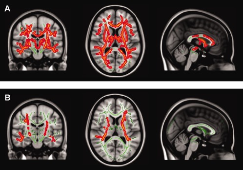
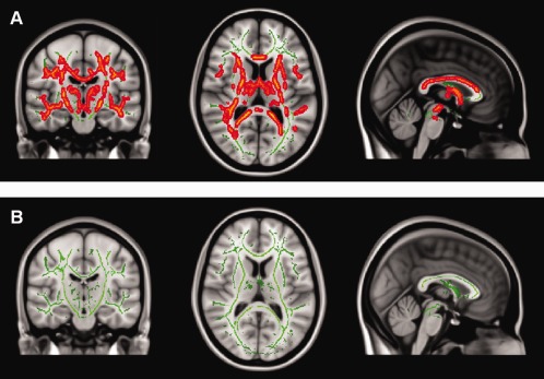
Results: Widespread cortical and deep WM area differences characterized by increased FA values were seen in the AOAMS compared with POMS group (P < 0.05, TFCE corrected). Significantly increased FA values of posterior WM areas were detected in the AODMS compared with POMS group (P < 0.05, TFCE corrected).
Conclusion: Increased FA values in WM areas of the AOMS compared with the POMS patients suggest that diffuse WM microstructure changes are more attributable to age of onset than a simple function of disease duration and age.
Keywords: adult-onset; diffusion-tensor-imaging; multiple sclerosis; pediatric-onset; tract-based spatial statistics (TBSS).
Copyright © 2012 Wiley Periodicals, Inc.
Figures
References
-
- Ashburner J, Friston KJ (2000): Voxel‐based morphometry—The methods. Neuroimage 11(Part 1):805–821. - PubMed
-
- Banwell B, Shroff M, Ness JM, Jeffery D, Schwid S, Weinstock‐Guttman B (2007): MRI features of pediatric multiple sclerosis. Neurology 68(Suppl 2):S46–S53. - PubMed
-
- Barkhof F (1999): MRI in multiple sclerosis: Correlation with expanded disability status scale (EDSS). Mult Scler 5:283–286. - PubMed
-
- Bethune A, Tipu V, Sled JG, Narayanan S, Arnold DL, et al. (2011): Diffusion tensor imaging and cognitive speed in children with multiple sclerosis. J Neurol Sci 309:68–74. - PubMed
-
- Chabas D, Castillo‐Trivino T, Mowry EM, Strober JB, Glenn OA, Waubant E (2008a): Vanishing MS T2‐bright lesions before puberty: A distinct MRI phenotype? Neurology 71:1090–1093. - PubMed
MeSH terms
LinkOut - more resources
Full Text Sources
