. 2012 Aug 31;337(6098):1104-7.
doi: 10.1126/science.1219875.

Synthesis of methylphosphonic acid by marine microbes: a source for methane in the aerobic ocean

Affiliations

Synthesis of methylphosphonic acid by marine microbes: a source for methane in the aerobic ocean

William W Metcalf et al. Science. .

Abstract

Relative to the atmosphere, much of the aerobic ocean is supersaturated with methane; however, the source of this important greenhouse gas remains enigmatic. Catabolism of methylphosphonic acid by phosphorus-starved marine microbes, with concomitant release of methane, has been suggested to explain this phenomenon, yet methylphosphonate is not a known natural product, nor has it been detected in natural systems. Further, its synthesis from known natural products would require unknown biochemistry. Here we show that the marine archaeon Nitrosopumilus maritimus encodes a pathway for methylphosphonate biosynthesis and that it produces cell-associated methylphosphonate esters. The abundance of a key gene in this pathway in metagenomic data sets suggests that methylphosphonate biosynthesis is relatively common in marine microbes, providing a plausible explanation for the methane paradox.


Figures
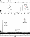
Fig. 1
Fig. 1
In vitro assay of MpnS activity. (A) Crude cell extract from an E. coli MpnS overexpression strain was incubated aerobically with 1-13C-HEP in the presence of Fe(II) and the phosphorus-containing products were examined using 31P NMR spectroscopy. After incubation for 1 hour a single product was observed as a doublet centered at 23.5 ppm. The mass and retention time of this product determined by LC-MS is consistent with this product being 1-13CMPn (Fig. S3). (B) Spiking of this reaction with the substrate, 1-13C-HEP produced a second doublet centered at 19 ppm, showing that the substrate was completely consumed in the initial reaction. (C) The identity of the reaction products was determined using 13C NMR after repeating the assay in a sealed vial using purified MpnS with a mixture of 1-13C-HEP and 2-13C-HEP as substrates. The C-2 labeled carbon of HEP is converted to 13C bicarbonate (H13CO -3), while the C-1-labelled carbon is converted to 1-13C-MPn. Bonding to phosphorus splits the 13C peak in the NMR spectrum. Thus, the C-1 peak is split and the C-2 peak is not. Glycerol, a component of the assay mixture, is also observed in the 13C spectrum. The 13C label is indicated by a red asterisk.
Fig. 2
Fig. 2
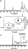
In vivo production of methylphosphonate esters by N. maritimus. (A) A cell extract of N. maritimus was prepared by sonication of whole cells as described. After removal of the cell debris by centrifugation, the supernatant was examined by 31P NMR spectroscopy revealing at least two compounds with chemical shifts in the range typical of phosphonic acids. (B) The two-dimensional HMBC NMR spectrum of N. maritimus cell extract. Comparison of the proton splitting patterns (shown in the insets) to those of model compounds (Figs S6 & S7) clearly shows that the P compound at 28 ppm in the 31P dimension is a methylphosphonate ester. The doublet of the proton at 1.4 ppm coupled to the phosphorus is diagnostic for a methyl group bonded directly to phosphorus, i.e. a methylphosphonate moiety. (C) High resolution LC-MS analysis showing the presence of free methylphosphonate after strong acid hydrolysis of N. maritimus cell debris. The extracted ion chromatogram centered around m/z 94.99035 (exact monoisotopic mass of methylphosphonate [M-H]-) with Fourier-transform mass spectrum and ion structure is shown in the inset. The chromatographic and MS fragmentation pattern is identical to an authentic MPn standard (Fig S8).
Fig. 3
Fig. 3
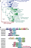
(A) The evolutionary relationships of biochemically characterized MpnS, HepD and HppE proteins (shown in bold) and homologs recovered from Genbank and the GOS metagenomic dataset was inferred using maximum likelihood analysis as described. Bootstrap values from 100 replicates are shown at the nodes. Robust bootstrap support for the tree shows that the method clearly differentiates MpnS (green), HepD (blue) and HppE (red) proteins. The full tree with all individual homologs shown is presented in Fig S9. (B) The gene content of large scaffolds containing the GOS MpnS homologs is compared to the mpnS locus of N. maritimus. The grey boxes represent sequencing gaps between paired-end reads.

Similar articles

Cited by

References

    1. Rogers JE, Whitman WB, editors. Microbial production and consumption of greenhouse gases : methane, nitrogen oxides, and halomethanes. American Society for Microbiology; Washington, D.C.: 1991.
    1. Karl DM, et al. Nat. Geosci. 2008;1:473.
    1. Reeburgh WS. Chem. Rev. 2007;107:486. - PubMed
    1. Daughton CG, Cook AM, Alexander M. FEMS Microbiol. Lett. 1979;5:91.
    1. Martinez A, Tyson GW, DeLong EF. Environ. Microbiol. 2010;12:222. - PubMed

Publication types