. 2012 Oct 12;287(42):34927-34935.
doi: 10.1074/jbc.M112.376814. Epub 2012 Aug 30.

Transmembrane and extracellular domains of syndecan-1 have distinct functions in regulating lung epithelial migration and adhesion

Affiliations

Transmembrane and extracellular domains of syndecan-1 have distinct functions in regulating lung epithelial migration and adhesion

William A Altemeier et al. J Biol Chem. .

Abstract

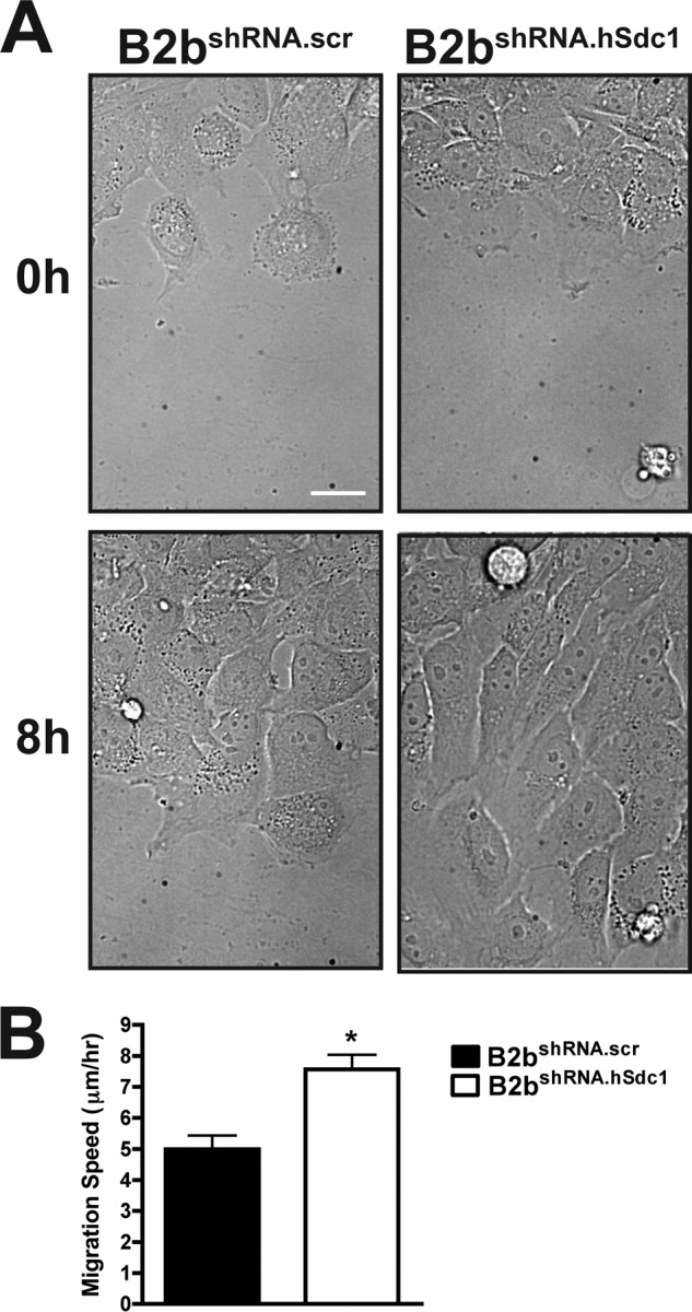
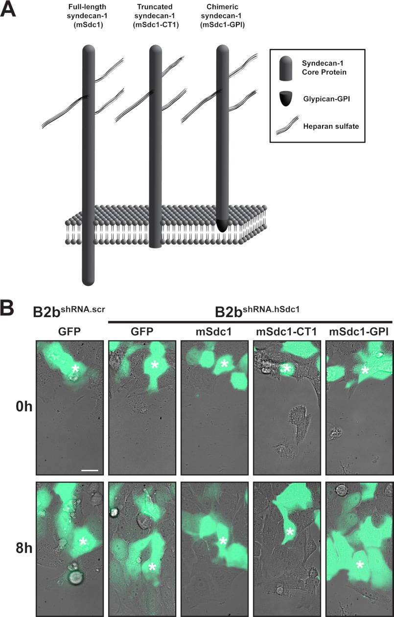
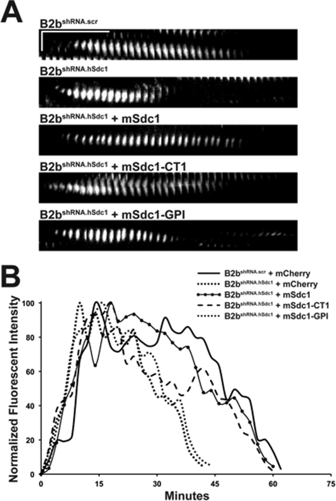
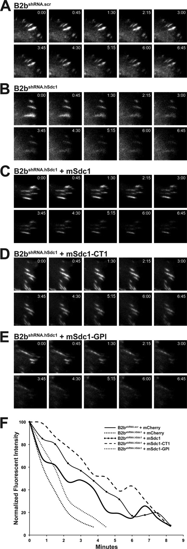
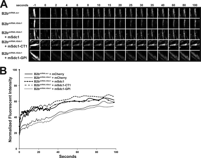
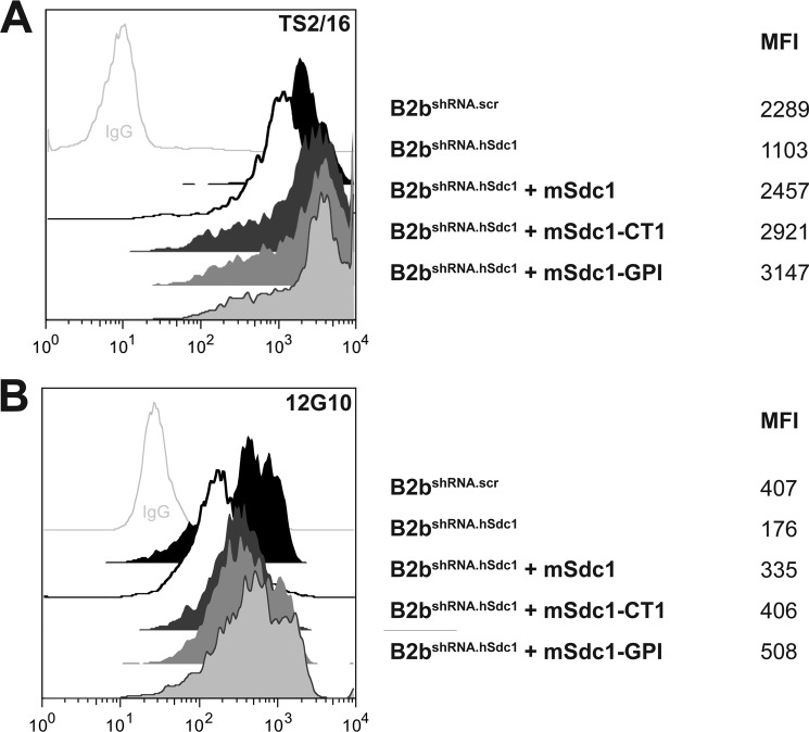
Syndecan-1 is a cell surface proteoglycan that can organize co-receptors into a multimeric complex to transduce intracellular signals. The syndecan-1 core protein has multiple domains that confer distinct cell- and tissue-specific functions. Indeed, the extracellular, transmembrane, and cytoplasmic domains have all been found to regulate specific cellular processes. Our previous work demonstrated that syndecan-1 controls lung epithelial migration and adhesion. Here, we identified the necessary domains of the syndecan-1 core protein that modulate its function in lung epithelial repair. We found that the syndecan-1 transmembrane domain has a regulatory function in controlling focal adhesion disassembly, which in turn controls cell migration speed. In contrast, the extracellular domain facilitates cell adhesion through affinity modulation of α(2)β(1) integrin. These findings highlight the fact that syndecan-1 is a multidimensional cell surface receptor that has several regulatory domains to control various biological processes. In particular, the lung epithelium requires the syndecan-1 transmembrane domain to govern cell migration and is independent from its ability to control cell adhesion via the extracellular domain.


Figures

FIGURE 1.
FIGURE 1.
Syndecan-1 restrains cell migration. A, monolayers of B2bshRNA.scr and B2bshRNA.hSdc1 cells were injured, and the wound closure was observed over time. See supplemental Video 1 for the entire image series. Scale bar = 20 μm. B, migration speed was measured for cells at the injury front. *, p < 0.0005 by Student's t test (n = four independent experiments). Migration speed for a minimum of 10 cells was measured for each condition in each experiment.
FIGURE 2.
FIGURE 2.
Transmembrane domain of syndecan-1 regulates migration speed. A, schematic of wild-type and mutant syndecan-1 used in these experiments. B, B2bshRNA.scr and B2bshRNA.hSdc1 cells transduced as indicated were injured, and migration of eGFP cells was observed. White asterisks identify the same cell at 0 and 8 h after injury. See supplemental Video 2 for the entire image series. Scale bar = 20 μm.
FIGURE 3.
FIGURE 3.
Syndecan-1 transmembrane domain slows focal adhesion disassembly in migrating lung epithelial cells. Cells stably expressing paxillin-eGFP were injured, and the turnover of focal adhesions in migrating cells was evaluated. A, kymographs of a focal adhesion at the leading edge of a migrating cell. Horizontal line = 10 min; vertical line = 5 μm. B, the normalized intensity of the focal adhesion in A was measured and plotted over time.
FIGURE 4.
FIGURE 4.
Syndecan-1 transmembrane domain attenuates focal adhesion disassembly after nocodazole washout. Cells stably expressing paxillin-eGFP were treated with nocodazole (10 μm, 4 h), and the disassembly of focal adhesions was evaluated after washout. A–E, focal adhesion disassembly was observed in B2bshRNA.scr cells, B2bshRNA.hSdc1 cells, and B2bshRNA.hSdc1 cells expressing wild-type and mutant mouse syndecan-1. F, intensity curves of the focal adhesions in A–E.
FIGURE 5.
FIGURE 5.
Syndecan-1 transmembrane domain facilitates recovery of fluorescence after photobleaching in migrating lung epithelial cells. Cells stably expressing paxillin-eGFP were injured, and the focal adhesion at the leading edge of the migrating cells was selectively photobleached. A, focal adhesion prior to (−1 s) and after photobleaching. B, normalized intensity of the representative focal adhesion in A plotted over time.
FIGURE 6.
FIGURE 6.
Syndecan-1 ectodomain regulates activation of β1 integrin subunit. Shown are representative histograms of the high affinity β1 integrin subunit using antibody clones TS2/16 (A) and 12G10 (B). The cell line and geometric mean fluorescence intensity (MFI) are indicated to the right of the respective histogram.

Similar articles

Cited by

References

    1. Couchman J. R. (2003) Syndecans: proteoglycan regulators of cell-surface microdomains? Nat. Rev. Mol. Cell Biol. 4, 926–937 - PubMed
    1. Teng Y. H., Aquino R. S., Park P. W. (2012) Molecular functions of syndecan-1 in disease. Matrix Biol. 31, 3–16 - PMC - PubMed
    1. Couchman J. R. (2010) Transmembrane signaling proteoglycans. Annu. Rev. Cell Dev. Biol. 26, 89–114 - PubMed
    1. Beauvais D. M., Rapraeger A. C. (2003) Syndecan-1-mediated cell spreading requires signaling by αvβ3 integrins in human breast carcinoma cells. Exp. Cell Res. 286, 219–232 - PubMed
    1. Beauvais D. M., Burbach B. J., Rapraeger A. C. (2004) The syndecan-1 ectodomain regulates αvβ3 integrin activity in human mammary carcinoma cells. J. Cell Biol. 167, 171–181 - PMC - PubMed

Publication types