

Isolating a cerebellar contribution to rapid visual attention using transcranial magnetic stimulation
- PMID: 22936903
- PMCID: PMC3426766
- DOI: 10.3389/fnbeh.2012.00055
Isolating a cerebellar contribution to rapid visual attention using transcranial magnetic stimulation
Abstract
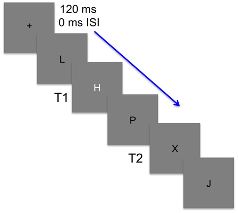
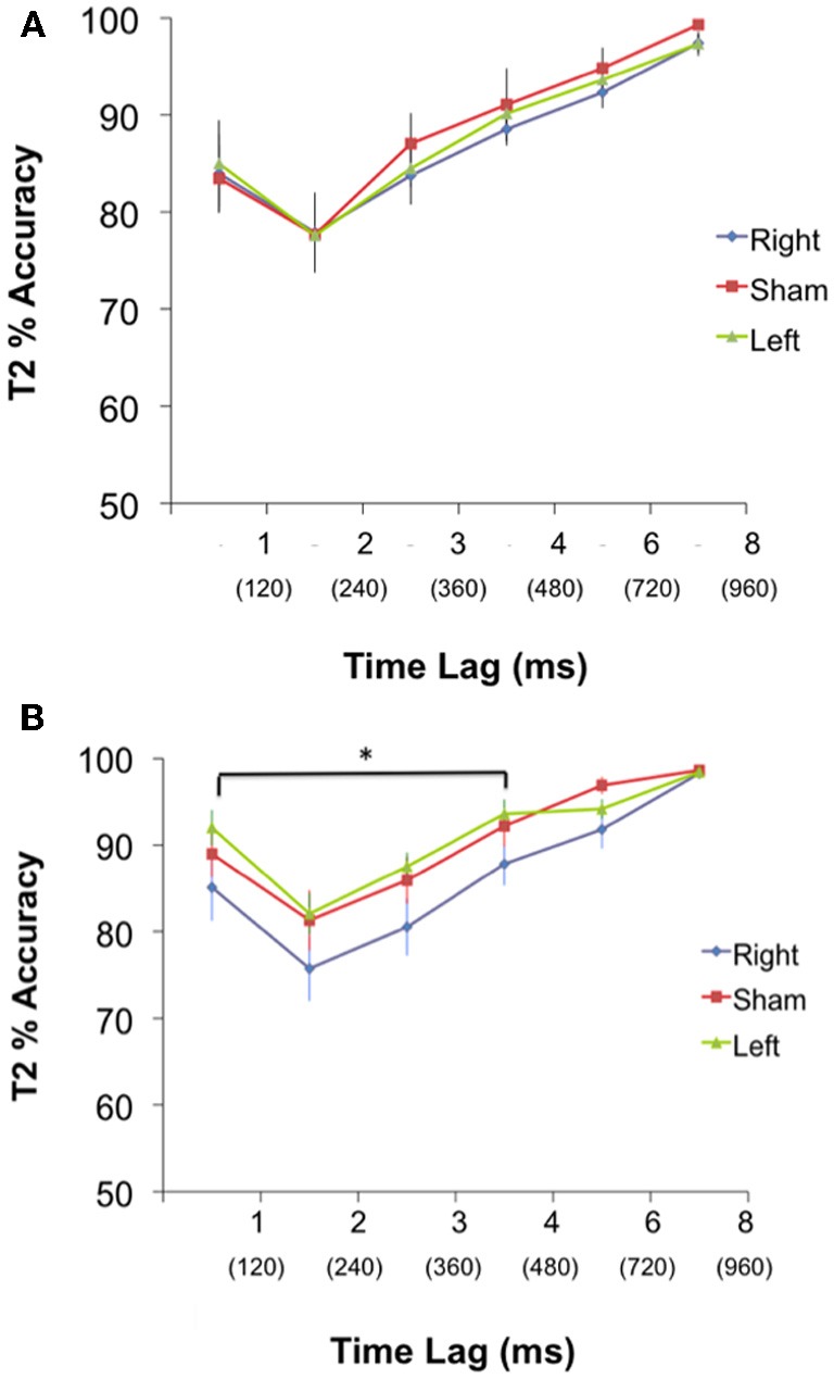
Patient and neuroimaging research have provided increasing support for a role of the posterior-lateral cerebellum in cognition, particularly attention. During rapid serial visual presentation, when two targets are presented in close temporal proximity (<500 ms), accuracy at detecting the second target (T2) suffers. This phenomenon is known as the attentional blink (AB), and in cerebellar lesion patients this effect is exaggerated. Damage to the cerebellum may thus disrupt the use of attentional resources during stimulus processing conditions that are temporally demanding. There are reciprocal connections between the cerebral cortex and the contralateral cerebellum, these connections allow for the possibility that lateralized functions in the cerebral cortex (such as language) remain lateralized in the cerebellum. The purpose of this study was to investigate the temporal characteristics of the cerebellar contribution to the AB and to functionally localize the contribution of the cerebellum to the AB using transcranial magnetic stimulation (TMS). We hypothesized that T2 accuracy would decrease after right cerebellar stimulation when the delay between the first target (T1) and T2 was short (120-400 ms) compared to long (720-960 ms). We used continuous theta burst stimulation (cTBS), a form of TMS, to transiently inhibit a focal population of neurons in the left and right posterior-lateral cerebellum of healthy participants (n = 45). Three groups of participants (n = 15) performed the AB before and after either sham, left, or right cerebellar stimulation. The results of this cTBS study support our hypothesis. During the short delay, participants in the right cTBS group showed a greater AB magnitude compared to both the left and sham cTBS groups (p < 0.05). No difference in T2 detection was found over long delays. The results provide further support for a cerebellar contribution to an integrated neural network recruited during temporally demanding attention-based tasks.
Keywords: attentional blink; cerebellum; cognition; theta burst stimulation; visual attention.
Figures
Similar articles
-
Modulation of cognitive cerebello-cerebral functional connectivity by lateral cerebellar continuous theta burst stimulation.Neuroimage. 2017 Sep;158:48-57. doi: 10.1016/j.neuroimage.2017.06.048. Epub 2017 Jun 29. Neuroimage. 2017. PMID: 28669908
-
Both 50 and 30 Hz continuous theta burst transcranial magnetic stimulation depresses the cerebellum.Cerebellum. 2019 Apr;18(2):157-165. doi: 10.1007/s12311-018-0971-0. Cerebellum. 2019. PMID: 30117122 Clinical Trial.
-
Long-lasting inhibition of cerebellar output.Brain Stimul. 2010 Jul;3(3):161-9. doi: 10.1016/j.brs.2009.10.001. Epub 2009 Oct 31. Brain Stimul. 2010. PMID: 20633445
-
Safety Considerations for Cerebellar Theta Burst Stimulation.Clin Ther. 2020 Jul;42(7):1169-1190.e1. doi: 10.1016/j.clinthera.2020.06.001. Epub 2020 Jul 14. Clin Ther. 2020. PMID: 32674957
-
Timing Tasks Synchronize Cerebellar and Frontal Ramping Activity and Theta Oscillations: Implications for Cerebellar Stimulation in Diseases of Impaired Cognition.Front Psychiatry. 2016 Jan 18;6:190. doi: 10.3389/fpsyt.2015.00190. eCollection 2015. Front Psychiatry. 2016. PMID: 26834650 Free PMC article. Review.
Cited by
-
Probing cerebellar involvement in cognition through a meta-analysis of TMS evidence.Sci Rep. 2021 Jul 20;11(1):14777. doi: 10.1038/s41598-021-94051-5. Sci Rep. 2021. PMID: 34285287 Free PMC article.
-
Network-targeted cerebellar transcranial magnetic stimulation improves attentional control.Neuroimage. 2017 Aug 1;156:190-198. doi: 10.1016/j.neuroimage.2017.05.011. Epub 2017 May 8. Neuroimage. 2017. PMID: 28495634 Free PMC article.
-
Targeting the Cerebellum by Noninvasive Neurostimulation: a Review.Cerebellum. 2017 Jun;16(3):695-741. doi: 10.1007/s12311-016-0840-7. Cerebellum. 2017. PMID: 28032321 Review.
-
The Cerebellum Modulates Attention Network Functioning: Evidence from a Cerebellar Transcranial Direct Current Stimulation and Attention Network Test Study.Cerebellum. 2019 Jun;18(3):457-468. doi: 10.1007/s12311-019-01014-8. Cerebellum. 2019. PMID: 30798474 Clinical Trial.
-
Effects of tDCS on the attentional blink revisited: A statistical evaluation of a replication attempt.PLoS One. 2022 Jan 27;17(1):e0262718. doi: 10.1371/journal.pone.0262718. eCollection 2022. PLoS One. 2022. PMID: 35085301 Free PMC article.
References
-
- Appollonio I. M., Grafman J., Schwartz V., Massaquoi S., Hallett M. (1993). Memory in patients with cerebellar degeneration. Neurology 43, 1536–1544 - PubMed
-
- Broadbent D. E., Broadbent M. H. (1987). From detection to identification: response to multiple targets in rapid serial visual presentation. Percept. Psychophys. 42, 105–113 - PubMed
LinkOut - more resources
Full Text Sources
Miscellaneous