

Emotional and cognitive processing of narratives and individual appraisal styles: recruitment of cognitive control networks vs. modulation of deactivations
- PMID: 22936905
- PMCID: PMC3427542
- DOI: 10.3389/fnhum.2012.00239
Emotional and cognitive processing of narratives and individual appraisal styles: recruitment of cognitive control networks vs. modulation of deactivations
Abstract
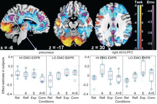
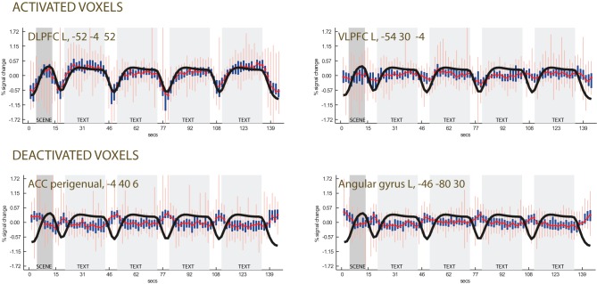
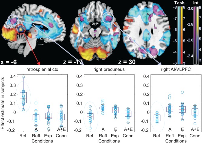
Research in psychotherapy has shown that the frequency of use of specific classes of words (such as terms with emotional valence) in descriptions of scenes of affective relevance is a possible indicator of psychological affective functioning. Using functional magnetic resonance imaging (MRI), we investigated the neural correlates of these linguistic markers in narrative texts depicting core aspects of emotional experience in human interaction, and their modulation by individual differences in the propensity to use these markers. Emotional words activated both lateral and medial aspects of the prefrontal cortex, as in previous studies of instructed emotion regulation and in consistence with recruitment of effortful control processes. However, individual differences in the spontaneous use of emotional terms in characterizing the stimulus material were prevalently associated with modulation of the signal in the perigenual cortex, in the retrosplenial cortex and precuneus, and the anterior insula/ventrolateral prefrontal cortex. Modulation of signal by the presence of these textual markers or individual differences mostly involved areas deactivated by the main task, thus further differentiating neural correlates of these appraisal styles from those associated with effortful control. These findings are discussed in the context of reports in the literature of modulations of deactivations, which suggest their importance in orienting attention and generation of response in the presence of emotional information. These findings suggest that deactivations may play a functional role in emotional appraisal and may contribute to characterizing different appraisal styles.
Keywords: appraisal; appraisal styles; emotion; reading; self-regulation.
Figures
Similar articles
-
Dysfunctional modulation of emotional interference in the medial prefrontal cortex in patients with schizophrenia.Neurosci Lett. 2008 Aug 1;440(2):119-24. doi: 10.1016/j.neulet.2008.05.094. Epub 2008 Jun 16. Neurosci Lett. 2008. PMID: 18562102 Clinical Trial.
-
Investigating the Neural Correlates of Emotion-Cognition Interaction Using an Affective Stroop Task.Front Psychol. 2017 Sep 1;8:1489. doi: 10.3389/fpsyg.2017.01489. eCollection 2017. Front Psychol. 2017. PMID: 28919871 Free PMC article.
-
Neural circuitry of emotion regulation: Effects of appraisal, attention, and cortisol administration.Cogn Affect Behav Neurosci. 2017 Apr;17(2):437-451. doi: 10.3758/s13415-016-0489-1. Cogn Affect Behav Neurosci. 2017. PMID: 28032303 Clinical Trial.
-
Neural Circuitry of Impaired Emotion Regulation in Substance Use Disorders.Am J Psychiatry. 2016 Apr 1;173(4):344-61. doi: 10.1176/appi.ajp.2015.15060710. Epub 2016 Jan 15. Am J Psychiatry. 2016. PMID: 26771738 Free PMC article. Review.
-
The neural basis of one's own conscious and unconscious emotional states.Neurosci Biobehav Rev. 2015 Oct;57:1-29. doi: 10.1016/j.neubiorev.2015.08.003. Epub 2015 Aug 4. Neurosci Biobehav Rev. 2015. PMID: 26363579 Review.
Cited by
-
Second Language Use Facilitates Implicit Emotion Regulation via Content Labeling.Front Psychol. 2017 Mar 16;8:366. doi: 10.3389/fpsyg.2017.00366. eCollection 2017. Front Psychol. 2017. PMID: 28360873 Free PMC article.
-
Changing views of emotion regulation and neurobiological models of the mechanism of action of psychotherapy.Cogn Affect Behav Neurosci. 2016 Aug;16(4):571-87. doi: 10.3758/s13415-016-0440-5. Cogn Affect Behav Neurosci. 2016. PMID: 27351671 Review.
-
Prestige and content biases together shape the cultural transmission of narratives.Evol Hum Sci. 2021 Jul 29;3:e42. doi: 10.1017/ehs.2021.37. eCollection 2021. Evol Hum Sci. 2021. PMID: 37588523 Free PMC article.
-
Abnormal Default System Functioning in Depression: Implications for Emotion Regulation.Front Psychol. 2016 Jun 10;7:858. doi: 10.3389/fpsyg.2016.00858. eCollection 2016. Front Psychol. 2016. PMID: 27375536 Free PMC article. Review.
-
Emotion regulation, attention to emotion, and the ventral attentional network.Front Hum Neurosci. 2013 Nov 7;7:746. doi: 10.3389/fnhum.2013.00746. Front Hum Neurosci. 2013. PMID: 24223546 Free PMC article. Review.
References
-
- Ainsworth M. D. S., Blear M. C., Waters E., Wail S. (1978). Patterns of Attachment: A Psychological Study of the Strange Situation. Hillsdale, IL: Erlbaum
LinkOut - more resources
Full Text Sources