

Resting-state connectivity of the amygdala is altered following Pavlovian fear conditioning
- PMID: 22936906
- PMCID: PMC3426015
- DOI: 10.3389/fnhum.2012.00242
Resting-state connectivity of the amygdala is altered following Pavlovian fear conditioning
Abstract
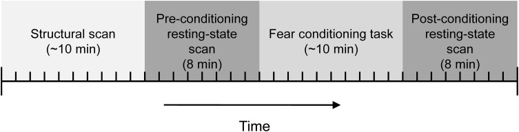
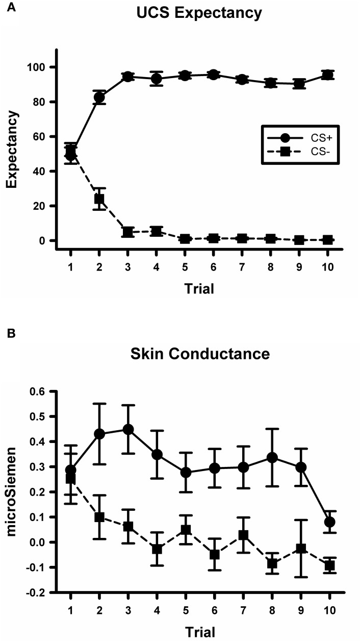
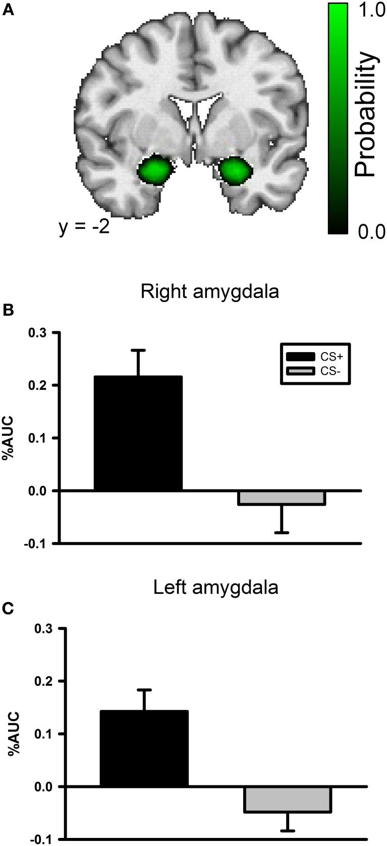
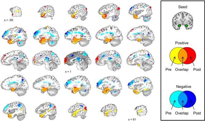
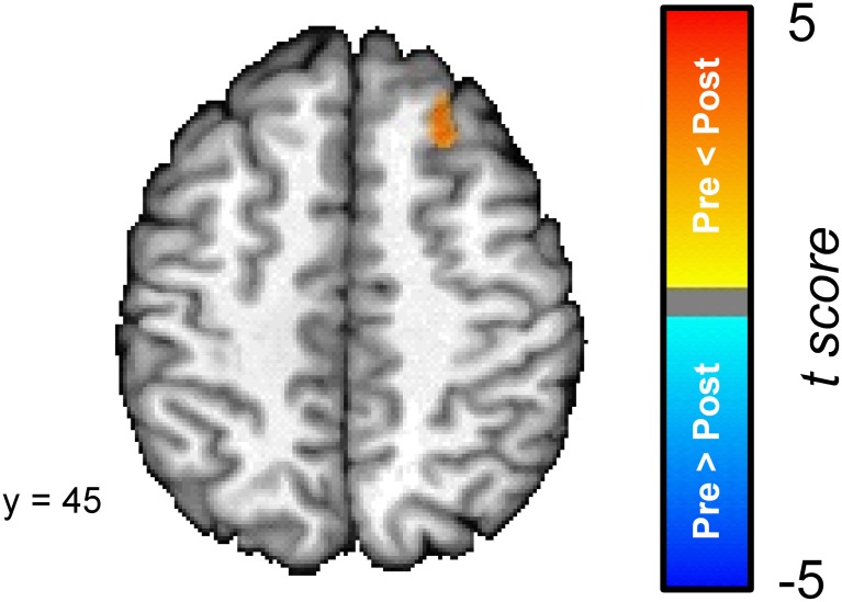
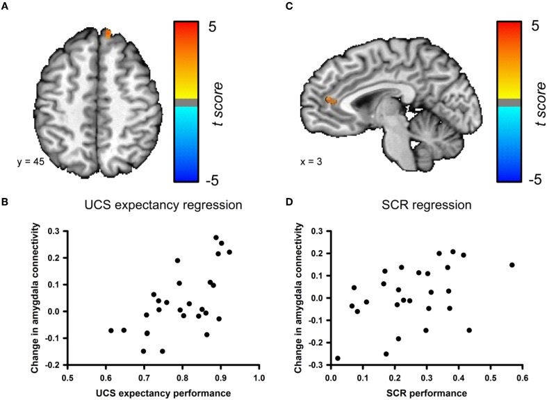
Neural plasticity in the amygdala is necessary for the acquisition and storage of memory in Pavlovian fear conditioning, but most neuroimaging studies have focused only on stimulus-evoked responses during the conditioning session. This study examined changes in the resting-state functional connectivity (RSFC) of the amygdala before and after Pavlovian fear conditioning, an emotional learning task. Behavioral results from the conditioning session revealed that participants learned normally and fMRI data recorded during learning identified a number of stimulus-evoked changes that were consistent with previous work. A direct comparison between the pre- and post-conditioning amygdala connectivity revealed a region of dorsal prefrontal cortex (PFC) in the superior frontal gyrus that showed a significant increase in connectivity following the conditioning session. A behavioral measure of explicit memory performance was positively correlated with the change in amygdala connectivity within a neighboring region in the superior frontal gyrus. Additionally, an implicit autonomic measure of conditioning was positively correlated with the change in connectivity between the amygdala and the anterior cingulate cortex (ACC). The resting-state data show that amygdala connectivity is altered following Pavlovian fear conditioning and that these changes are also related to behavioral outcomes. These alterations may reflect the operation of a consolidation process that strengthens neural connections to support memory after the learning event.
Keywords: amygdala; fMRI; fear conditioning; functional connectivity; resting-state.
Figures
Similar articles
-
Memory consolidation of fear conditioning: bi-stable amygdala connectivity with dorsal anterior cingulate and medial prefrontal cortex.Soc Cogn Affect Neurosci. 2014 Nov;9(11):1730-7. doi: 10.1093/scan/nst170. Epub 2013 Nov 4. Soc Cogn Affect Neurosci. 2014. PMID: 24194579 Free PMC article.
-
Longitudinal changes of resting-state functional connectivity of amygdala following fear learning and extinction.Int J Psychophysiol. 2020 Mar;149:15-24. doi: 10.1016/j.ijpsycho.2020.01.002. Epub 2020 Jan 9. Int J Psychophysiol. 2020. PMID: 31926905
-
Resting-state functional connectivity between amygdala and the ventromedial prefrontal cortex following fear reminder predicts fear extinction.Soc Cogn Affect Neurosci. 2016 Jun;11(6):991-1001. doi: 10.1093/scan/nsw031. Epub 2016 Mar 24. Soc Cogn Affect Neurosci. 2016. PMID: 27013104 Free PMC article.
-
Interplay of amygdala and cingulate plasticity in emotional fear.Neural Plast. 2011;2011:813749. doi: 10.1155/2011/813749. Epub 2011 Sep 7. Neural Plast. 2011. PMID: 21912749 Free PMC article. Review.
-
The amygdala and medial prefrontal cortex: partners in the fear circuit.J Physiol. 2013 May 15;591(10):2381-91. doi: 10.1113/jphysiol.2012.248575. Epub 2013 Feb 18. J Physiol. 2013. PMID: 23420655 Free PMC article. Review.
Cited by
-
From Pavlov to PTSD: the extinction of conditioned fear in rodents, humans, and anxiety disorders.Neurobiol Learn Mem. 2014 Sep;113:3-18. doi: 10.1016/j.nlm.2013.11.014. Epub 2013 Dec 7. Neurobiol Learn Mem. 2014. PMID: 24321650 Free PMC article. Review.
-
Threat bias and resting state functional connectivity of the amygdala and bed nucleus stria terminalis.J Psychiatr Res. 2020 Mar;122:54-63. doi: 10.1016/j.jpsychires.2019.12.017. Epub 2019 Dec 31. J Psychiatr Res. 2020. PMID: 31927266 Free PMC article.
-
An externally validated resting-state brain connectivity signature of pain-related learning.Commun Biol. 2024 Jul 17;7(1):875. doi: 10.1038/s42003-024-06574-y. Commun Biol. 2024. PMID: 39020002 Free PMC article.
-
Extended amygdala connectivity changes during sustained shock anticipation.Transl Psychiatry. 2018 Jan 31;8(1):33. doi: 10.1038/s41398-017-0074-6. Transl Psychiatry. 2018. PMID: 29382815 Free PMC article.
-
Threat of shock increases excitability and connectivity of the intraparietal sulcus.Elife. 2017 May 30;6:e23608. doi: 10.7554/eLife.23608. Elife. 2017. PMID: 28555565 Free PMC article.
References
-
- Biswal B., Mennes M., Zuo X., Gohel S., Kelly C., Smith S., Beckmann C., Adelstein J., Buckner R., Colcombe S., Dogonowski A., Ernst M., Fair D., Hampson M., Hoptman M., Hyde J., Kiviniemi V., Kotter R., Li S., Lin C., Lowe M., Mackay C., Madden D., Madsen K., Margulies D., Mayberg H., McMahon K., Monk C., Mostofsky S., Nagel B., Pekar J., Peltier S., Petersen S., Riedl V., Rombouts S., Rypma B., Schlaggar B., Schmidt S., Seidler R., Siegle G., Sorg C., Teng G., Veijola J., Villringer A., Walter M., Wang L., Weng X., Whitfield-Gabrieli S., Williamson P., Windischberger C., Zang Y., Zhang H., Castellanos F., Milham M. (2010). Toward discovery science of human brain function. Proc. Natl. Acad. Sci. U.S.A. 107, 4734–4739 10.1073/pnas.0911855107 - DOI - PMC - PubMed
-
- Cheng D., Knight D., Smith C., Stein E., Helmstetter F. (2003). Functional MRI of human amygdala activity during Pavlovian fear conditioning: stimulus processing versus response expression. Behav. Neurosci. 117, 3–10 - PubMed
Grants and funding
LinkOut - more resources
Full Text Sources
Miscellaneous