. 2012 Aug 22:3:204.
doi: 10.3389/fpls.2012.00204. eCollection 2012.

Comparative structure and biomechanics of plant primary and secondary cell walls

Affiliations

Comparative structure and biomechanics of plant primary and secondary cell walls

Daniel J Cosgrove et al. Front Plant Sci. .

Abstract

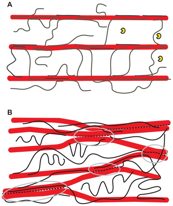
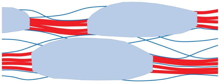
Recent insights into the physical biology of plant cell walls are reviewed, summarizing the essential differences between primary and secondary cell walls and identifying crucial gaps in our knowledge of their structure and biomechanics. Unexpected parallels are identified between the mechanism of expansion of primary cell walls during growth and the mechanisms by which hydrated wood deforms under external tension. There is a particular need to revise current "cartoons" of plant cell walls to be more consistent with data from diverse approaches and to go beyond summarizing limited aspects of cell walls, serving instead as guides for future experiments and for the application of new techniques.

Keywords: cellulose; creep; deformation; growth; primary cell walls; secondary cell walls; xyloglucan.


Figures

FIGURE 1
FIGURE 1
Alternative hypothetical architectures of cellulose– xyloglucan networks in primary cell walls. (A) The tethered network model in which xyloglucans (black lines) fully coat the surfaces of cellulose microfibrils (larger red rods) and additionally span the 20–40 nm gap between adjacent cellulose microfibrils as load-bearing tethers. The distance between microfibrils is large enough to permit penetration of a xyloglucanase-specific endoglucanase (yellow “Pacman” symbols) of the type used by Park and Cosgrove (2012a). This model is based a variety of results (Scheller and Ulvskov, 2010; Hayashi and Kaida, 2011) but is still hypothetical and recent work is at odds with it. In this model, the primary means of cell wall expansion is by cutting the xyloglucan tethers or by destabilizing the xyloglucan–cellulose interaction, resulting in wall stress relaxation and yielding. The directionality of growth depends on the net orientation of cellulose microfibrils (Baskin, 2005; Kerstens et al., 2001). (B) A revised architecture based on the enzyme/biomechanics analysis of Park and Cosgrove (2012a), in which the load-bearing xyloglucan (broken black lines highlighted by gray ellipses) is a minor fraction of the total xyloglucan and is trapped between microfibrils, so it is not accessible to xyloglucan-specific endoglucanase. By this arrangement the xyloglucan glues microfibrils into a network of microfibril bundles which serve to protect it from lytic action by xyloglucan-specific endoglucanases as well as xyloglucan endotransglycosylase/hydrolase. The gray circles demark the limited region that bear the static tensile forces generated by turgor pressure. An arrangement as shown here could account for the lack of wall loosening caused by xyloglucan endotransglycosylase/hydrolases (Saladie et al., 2006; Maris et al., 2009; Miedes et al., 2011).
FIGURE 2
FIGURE 2
Hypothetical architecture of polymer network in secondary cell walls of conifer wood. Loosely aggregated bundles of cellulose microfibrils (red) are coated with a disordered xylan–lignin complex (shaded light blue). Partially oriented glucomannan chains (blue lines) adhere by hydrogen bonding to the cellulose aggregates and acetylated segments of these glucomannans bridge between the aggregates. For clarity the structure is shown much more open than is the case: the free space, filled by water in vivo, is only about 40% of the total volume. This is much less than the free space in hydrated primary cell walls. Based on Terashima et al. (2009) and Fernandes et al. (2011).

References

    1. Abasolo W., Eder M., Yamauchi K., Obel N., Reinecke A., Neumetzler L., Dunlop J. W., Mouille G., Pauly M., Hofte H., Burgert I. (2009). Pectin may hinder the unfolding of xyloglucan chains during cell deformation: implications of the mechanical performance of Arabidopsis hypocotyls with pectin alterations. Mol. Plant 2 990–999 - PubMed
    1. Altaner C. M., Jarvis M. C. (2008). Modelling polymer interactions of the “molecular Velcro” type in wood under mechanical stress. J. Theor. Biol. 253 434–445 - PubMed
    1. Anderson C. T., Carroll A., Akhmetova L., Somerville C. (2010). Real-time imaging of cellulose reorientation during cell wall expansion in Arabidopsis roots. Plant Physiol. 152 787–796 - PMC - PubMed
    1. Assor C., Placet V., Chabbert B., Habrant A., Lapierre C., Pollet B., Perre P. (2009). Concomitant changes in viscoelastic properties and amorphous polymers during the hydrothermal treatment of hardwood and softwood. J. Agric. Food Chem. 57 6830–6837 - PubMed
    1. Atalla R. H., Hackney J. M., Uhlin I., Thompson N. S. (1993). Hemicelluloses as structure regulators in the aggregation of native cellulose. Int. J. Biol. Macromol. 15 109–112 - PubMed

LinkOut - more resources