The niche factor syndecan-1 regulates the maintenance and proliferation of neural progenitor cells during mammalian cortical development
- PMID: 22936997
- PMCID: PMC3427302
- DOI: 10.1371/journal.pone.0042883
The niche factor syndecan-1 regulates the maintenance and proliferation of neural progenitor cells during mammalian cortical development
Erratum in
- PLoS One. 2012;7(11). doi:10.1371/annotation/76cf3bd3-f842-489f-8ad2-4c016447b84c
Abstract
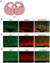
Neural progenitor cells (NPCs) divide and differentiate in a precisely regulated manner over time to achieve the remarkable expansion and assembly of the layered mammalian cerebral cortex. Both intrinsic signaling pathways and environmental factors control the behavior of NPCs during cortical development. Heparan sulphate proteoglycans (HSPG) are critical environmental regulators that help modulate and integrate environmental cues and downstream intracellular signals. Syndecan-1 (Sdc1), a major transmembrane HSPG, is highly enriched in the early neural germinal zone, but its function in modulating NPC behavior and cortical development has not been explored. In this study we investigate the expression pattern and function of Sdc1 in the developing mouse cerebral cortex. We found that Sdc1 is highly expressed by cortical NPCs. Knockdown of Sdc1 in vivo by in utero electroporation reduces NPC proliferation and causes their premature differentiation, corroborated in isolated cells in vitro. We found that Sdc1 knockdown leads to reduced levels of β-catenin, indicating reduced canonical Wnt signaling. Consistent with this, GSK3β inhibition helps rescue the Sdc1 knockdown phenotype, partially restoring NPC number and proliferation. Moreover, exogenous Wnt protein promotes cortical NPC proliferation, but this is prevented by Sdc1 knockdown. Thus, Sdc1 in the germinal niche is a key HSPG regulating the maintenance and proliferation of NPCs during cortical neurogenesis, in part by modulating the ability of NPCs to respond to Wnt ligands.
Conflict of interest statement
Figures


Similar articles
-
Syndecan-1 Promotes Streptococcus pneumoniae Corneal Infection by Facilitating the Assembly of Adhesive Fibronectin Fibrils.mBio. 2020 Dec 8;11(6):e01907-20. doi: 10.1128/mBio.01907-20. mBio. 2020. PMID: 33293379 Free PMC article.
-
Localisation of specific heparan sulfate proteoglycans during the proliferative phase of brain development.Dev Dyn. 2003 Jun;227(2):170-84. doi: 10.1002/dvdy.10298. Dev Dyn. 2003. PMID: 12761845
-
The Wnt/beta-catenin pathway directs neuronal differentiation of cortical neural precursor cells.Development. 2004 Jun;131(12):2791-801. doi: 10.1242/dev.01165. Epub 2004 May 13. Development. 2004. PMID: 15142975
-
Advances in the molecular functions of syndecan-1 (SDC1/CD138) in the pathogenesis of malignancies.Crit Rev Oncol Hematol. 2015 Apr;94(1):1-17. doi: 10.1016/j.critrevonc.2014.12.003. Epub 2014 Dec 18. Crit Rev Oncol Hematol. 2015. PMID: 25563413 Review.
-
Glycobiology of syndecan-1 in bacterial infections.Biochem Soc Trans. 2018 Apr 17;46(2):371-377. doi: 10.1042/BST20170395. Epub 2018 Mar 9. Biochem Soc Trans. 2018. PMID: 29523771 Free PMC article. Review.
Cited by
-
Electrical coupling regulates layer 1 interneuron microcircuit formation in the neocortex.Nat Commun. 2016 Aug 11;7:12229. doi: 10.1038/ncomms12229. Nat Commun. 2016. PMID: 27510304 Free PMC article.
-
Heparan Sulfate Proteoglycans: Key Mediators of Stem Cell Function.Front Cell Dev Biol. 2020 Nov 19;8:581213. doi: 10.3389/fcell.2020.581213. eCollection 2020. Front Cell Dev Biol. 2020. PMID: 33330458 Free PMC article. Review.
-
The Extracellular Matrix in the Evolution of Cortical Development and Folding.Front Cell Dev Biol. 2020 Dec 3;8:604448. doi: 10.3389/fcell.2020.604448. eCollection 2020. Front Cell Dev Biol. 2020. PMID: 33344456 Free PMC article. Review.
-
Quiescent horizontal basal stem cells act as a niche for olfactory neurogenesis in a mouse 3D organoid model.Cell Rep Methods. 2025 Jun 16;5(6):101055. doi: 10.1016/j.crmeth.2025.101055. Epub 2025 May 28. Cell Rep Methods. 2025. PMID: 40441150 Free PMC article.
-
The Control of Cortical Folding: Multiple Mechanisms, Multiple Models.Neuroscientist. 2024 Dec;30(6):704-722. doi: 10.1177/10738584231190839. Epub 2023 Aug 24. Neuroscientist. 2024. PMID: 37621149 Free PMC article. Review.
References
-
- Temple S (2001) The development of neural stem cells. Nature 414: 112–117. - PubMed
-
- Dehay C, Kennedy H (2007) Cell-cycle control and cortical development. Nat Rev Neurosci 8: 438–450. - PubMed
-
- Gotz M, Huttner WB (2005) The cell biology of neurogenesis. Nat Rev Mol Cell Biol 6: 777–788. - PubMed
-
- Noctor SC, Flint AC, Weissman TA, Dammerman RS, Kriegstein AR (2001) Neurons derived from radial glial cells establish radial units in neocortex. Nature 409: 714–720. - PubMed
-
- Buchman JJ, Tsai LH (2007) Spindle regulation in neural precursors of flies and mammals. Nat Rev Neurosci 8: 89–100. - PubMed
Publication types
MeSH terms
Substances
Grants and funding
LinkOut - more resources
Full Text Sources
Medical
Molecular Biology Databases
Research Materials
Miscellaneous

