. 2012;7(8):e43515.
doi: 10.1371/journal.pone.0043515. Epub 2012 Aug 24.

Proteomic analysis reveals new cardiac-specific dystrophin-associated proteins

Affiliations

Proteomic analysis reveals new cardiac-specific dystrophin-associated proteins

Eric K Johnson et al. PLoS One. 2012.

Abstract

Mutations affecting the expression of dystrophin result in progressive loss of skeletal muscle function and cardiomyopathy leading to early mortality. Interestingly, clinical studies revealed no correlation in disease severity or age of onset between cardiac and skeletal muscles, suggesting that dystrophin may play overlapping yet different roles in these two striated muscles. Since dystrophin serves as a structural and signaling scaffold, functional differences likely arise from tissue-specific protein interactions. To test this, we optimized a proteomics-based approach to purify, identify and compare the interactome of dystrophin between cardiac and skeletal muscles from as little as 50 mg of starting material. We found selective tissue-specific differences in the protein associations of cardiac and skeletal muscle full length dystrophin to syntrophins and dystrobrevins that couple dystrophin to signaling pathways. Importantly, we identified novel cardiac-specific interactions of dystrophin with proteins known to regulate cardiac contraction and to be involved in cardiac disease. Our approach overcomes a major challenge in the muscular dystrophy field of rapidly and consistently identifying bona fide dystrophin-interacting proteins in tissues. In addition, our findings support the existence of cardiac-specific functions of dystrophin and may guide studies into early triggers of cardiac disease in Duchenne and Becker muscular dystrophies.


Conflict of interest statement

Competing Interests: The authors have declared that no competing interests exist.

Figures

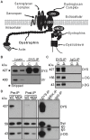
Figure 1
Figure 1. MANDYS1 specifically immunoprecipitates dystrophin and associated DAPC members.
(A) Schematic representation of the core DAPC in skeletal muscle. (B) MANDYS1 does not recognize utrophin. Western blot of lysates and DYS-IPs from cardiac (C) and skeletal (S) muscle probed for utrophin (Utr), then stripped and re-probed for dystrophin. (C) α- and β-dystroglycan are detected in DYS-IPs but not control IgG-IPs. (D) Syntrophins (Syn) and β-dystroglycan (β -DG) in DYS-IP from wild type but not mdx skeletal muscle or in IgG-IPs. Dystrophin is depleted in Post-IP lysates from WT muscle.
Figure 2
Figure 2. nNOS does not associate with full-length dystrophin in cardiomyocytes.
(A) Western blot analysis of DYS-IPs and IgG-IPs from wild type cardiac (C) and skeletal (S) muscle showing lack of nNOS detection in cardiac DYS-IP but presence of α1-syntrophin. (B) Immunolabeling of wild type (WT) and nNOS knock-out (nNOS−/−) cardiomyocytes for nNOS (green) and dystrophin (red) shows lack of co-localization. Arrows indicate non-specific labeling. (C) Peptide coverage (blue amino acids) by LC-MS/MS of spectrin repeats 16 and 17 of cardiac dystrophin.
Figure 3
Figure 3. Syntrophins differ between cardiac and skeletal muscle DAPC.
(A) Western blot analysis of syntrophins in mouse cardiac (C) and skeletal (S) muscle protein lysates, DYS-IPs and IgG-IPs. β2-syntrophin associates with dystrophin only in the heart. Fold differences in syntrophin abundance in cardiac vs. skeletal muscle DYS-IPs relative to dystrophin are shown (averages ±SD, N = 3). (B) Western blot analysis of DYS-IPs from human (H) and mouse (M) cardiac samples showing association of β2-syntrophin with dystrophin in the human heart. (C) Immunolabeling of cardiac sections from wild type mice for indicated proteins. Scale bar: 50 µm. Arrows indicate blood vessels.
Figure 4
Figure 4. Differences in α-dystrobrevin splice variants between cardiac and skeletal muscle DAPC.
(A) Western blot analysis of α-dystrobrevins in mouse cardiac (C) and skeletal (S) muscle total protein lysates, DYS-IPs and IgG-IPs. α3-dystrobrevin associates with dystrophin in the heart. Fold differences in α-dystrobrevin abundance in cardiac vs. skeletal muscle DYS-IPs relative to dystrophin are shown (averages ±SD, N = 3). (B) Immunolabeling of wild type cardiac sections for α1 and α2-dystrobrevins. Scale bar: 50 µm. (C) Immunolabeling of mdx cardiac tissue section for β-sarcoglycan. Scale bar: 50 µm. (D) Western blot analysis of α-dystrobrevins in heart protein lysates from wild type (WT) and mdx mice. Fold differences in α-dystrobrevin abundance in WT vs. mdx cardiac lysates relative to GAPDH are shown (averages ±SD, N = 3) (E) Western blot analysis of α-dystrobrevins in DYS-IPs from human (H) and mouse (M) cardiac samples. Additional α-dystrobrevin isoforms (arrow heads) are detected in human cardiac lysates and DYS-IP.
Figure 5
Figure 5. Novel cardiac-specific dystrophin-associated proteins.
(A) Western blot analysis of Cavin-1, Ahnak1, CRYAB, and Cypher in mouse cardiac and skeletal muscle lysates, DYS-IPs and IgG-IPs. (B) Cavin-1 and Ahnak1 co-purify with dystrophin in human cardiac samples. (C) Western blot analysis of Cavin-1 and Ahnak1 in MANDAG2-IPs from wild type (WT) and mdx cardiac muscle. Expression levels of Cavin-1 and Ahnak1 are not decreased in mdx lysates compared to WT.

References

    1. Ervasti JM, Sonnemann KJ (2008) Biology of the striated muscle dystrophin-glycoprotein complex. Int Rev Cytol 265: 191–225. - PubMed
    1. Albrecht DE, Froehner SC (2002) Syntrophins and dystrobrevins: defining the dystrophin scaffold at synapses. Neurosignals 11: 123–129. - PubMed
    1. Kaspar RW, Allen HD, Montanaro F (2009) Current understanding and management of dilated cardiomyopathy in Duchenne and Becker muscular dystrophy. J Am Acad Nurse Pract 21: 241–249. - PMC - PubMed
    1. Judge DP, Kass DA, Thompson WR, Wagner KR (2011) Pathophysiology and Therapy of Cardiac Dysfunction in Duchenne Muscular Dystrophy. Am J Cardiovasc Drugs. - PubMed
    1. Cohen N, Muntoni F (2004) Multiple pathogenetic mechanisms in X linked dilated cardiomyopathy. Heart 90: 835–841. - PMC - PubMed

Publication types