. 2012;7(8):e43593.
doi: 10.1371/journal.pone.0043593. Epub 2012 Aug 24.

Krüppel-like factor 4, a tumor suppressor in hepatocellular carcinoma cells reverts epithelial mesenchymal transition by suppressing slug expression

Affiliations

Krüppel-like factor 4, a tumor suppressor in hepatocellular carcinoma cells reverts epithelial mesenchymal transition by suppressing slug expression

Ze-Shiang Lin et al. PLoS One. 2012.

Erratum in

Abstract

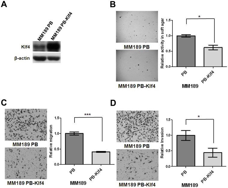
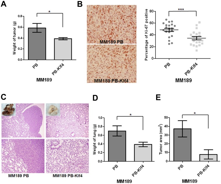
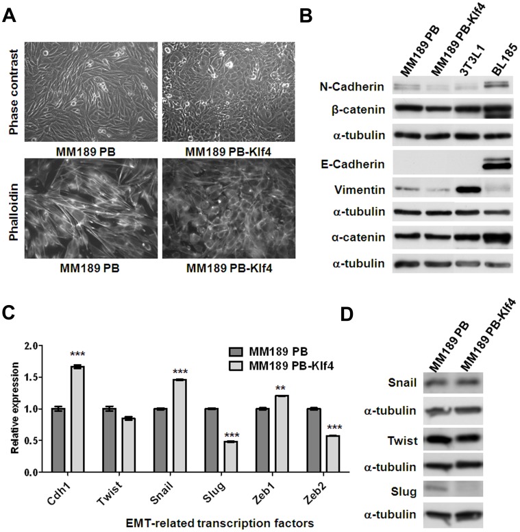
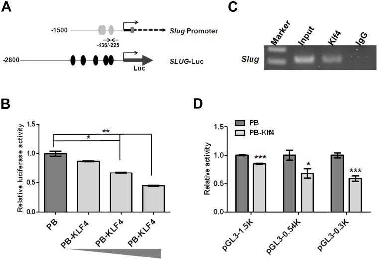
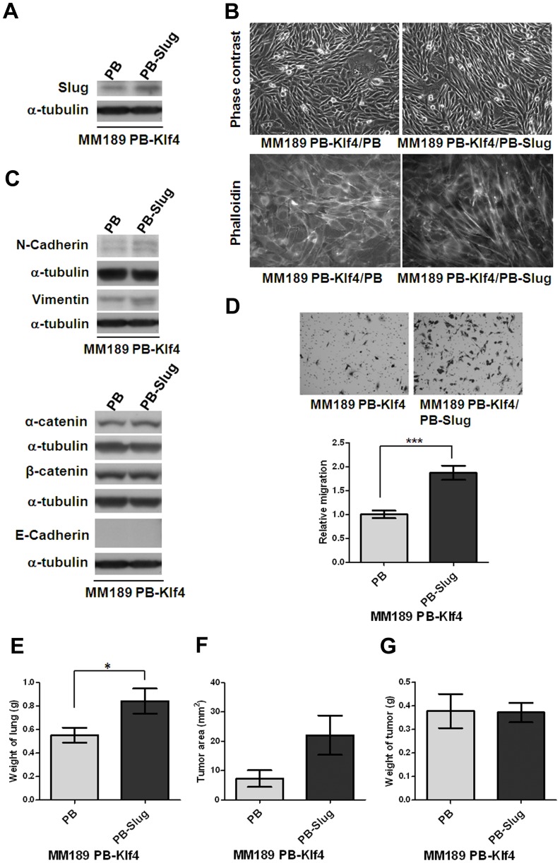
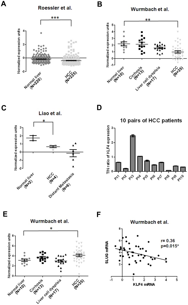
Krüppel-like factor 4 (KLF4) is a zinc-finger transcription factor that plays an important role in differentiation and pathogenesis. KLF4 has been suggested to act as an oncogene or tumor suppressor in different tumor types. However, the role of KLF4 in hepatocellular carcinoma (HCC) remains unclear. Here, we demonstrate that forced expression of Klf4 in murine HCC cell lines reduced anchorage-independent growth in soft agar as well as cell migration and invasion activities in vitro. Ectopic Klf4 expression impaired subcutaneous tumor growth and lung colonization in vivo. By contrast, Klf4 knockdown enhanced HCC cell migration. Interestingly, ectopic expression of Klf4 changed the morphology of murine HCC cells to a more epithelial phenotype. Associated with this, we found that expression of Slug, a critical epithelial mesenchymal transition (EMT)-related transcription factor, was significantly down-regulated in Klf4-expressing cells. Chromatin immunoprecipitation (ChIP) and luciferase reporter assays showed that Klf4 is able to bind and repress the activity of the Slug promoter. Furthermore, ectopic Slug expression partially reverts the Klf4-mediated phenotypes. Consistent with a role as a tumor suppressor in HCC, analysis of the public microarray databases from Oncomine revealed reduced KLF4 expression in human HCC tissues in comparison with normal liver tissues in 3 out of 4 data sets. By quantitative reverse transcription-polymerase chain reaction (qRT-PCR), we found reduced KLF4 mRNA in 50% of HCC tissues. Importantly, an inverse correlation between the expression of KLF4 and SLUG was found in HCC tissues. Our data suggest that KLF4 acts as a tumor suppressor in HCC cells, in part by suppressing SLUG transcription.


Conflict of interest statement

Competing Interests: The authors have declared that no competing interests exist.

Figures

Figure 1
Figure 1. Ectopic Klf4 expression inhibited colony formation, migration and invasion.
(A) Klf4 and β-actin protein levels were detected in murine HCC cell lines, MM189 with ectopic Klf4 expression (MM189 PB-Klf4) and its corresponding control (MM189 PB) by immunoblot assay. β-actin served as a loading control. (B) Representative anchorage-independent growth activity for MM189 cells with ectopic Klf4 expression (MM189 PB-Klf4) and its corresponding control (MM189 PB). The colonies were observed at lower magnification (40×) in the left panel. The relative activity was determined by normalizing the mean number of colonies in MM189 PB-Klf4 cells to that in MM189 PB cells. Bar, SE. *, p<0.05. (C) Representative data shows the relative migration activity of MM189 expressing Klf4 (MM189 PB-Klf4) and its vector control (MM189 PB). The migrated cells were observed at magnification (100×) in the left panel. The relative migration activity was defined by normalizing the mean of migrated cells/per field in MM189 PB-Klf4 cells to that in MM189 PB cells. Bar, SE. ***, p<0.001. (D) Representative data shows the relative invasion activity of MM189 expressing Klf4 (MM189 PB-Klf4) and its vector control (MM189 PB). The invaded cells were observed at magnification (100×) in the left panel. The relative invasion activity was defined by normalizing the mean of invaded cells/per field in MM189 PB-Klf4 cells to that in MM189 PB cells. Bar, SE. *, p<0.05.
Figure 2
Figure 2. Klf4 suppressed tumor growth and lung colonization.
(A) Quantification of the weight of the tumor lesions in mice (n = 7) subcutaneously injected with MM189 PB-Klf4 or MM189 PB cells. Bar, SE. *, p<0.05. (B) The representative field for detection of Ki-67 expression by immunohistochemistry under the light microscope with 200× magnification in the left panel. The percentage of positive Ki-67 stain was defined as the intensity of positive nuclei divided by that of the total nuclei in the field. Bar, SE. ***, p<0.001. (C) Representative lung fields of nude mice after the delivery, via tail vein injection, of MM189 cells with ectopic Klf4 expression (MM189 PB-Klf4) or vector controls (MM189 PB). The boxed area in the upper panel was shown with macroscopic view. The upper panel was observed at lower magnification (40×) and the lower was for higher magnification (100×). (D) Quantification of weight of the lung lesions in mice (n = 6) injected with MM189 PB-Klf4 or MM189 PB cells. Bar, SE. *, p<0.05. (E) Quantification of total areas of the tumor lesions in lungs of mice (n = 6) injected with MM189 PB-Klf4 or MM189 PB cells. Bar, SE. *, p<0.05.
Figure 3
Figure 3. Klf4 promoted an epithelial phenotype in MM189 cells.
(A) Ectopic Klf4 expression shifts cell morphology from a mesenchymal- to an epithelial phenotype. Phase contrast microscopy with 200× magnification (upper panel). Note the cobblestone appearance of the Klf4-expressing cells. Cytoskelton F-actin proteins were stained with rodamine-phalloidin and viewed under fluorescence microscope with 630× magnification (lower panel, shown in grey mode). (B) Immunoblot analysis of epithelial and mesenchymal proteins in MM189 PB and MM189 PB-Klf4 cells. BL185 cells and 3T3L1 cells served as positive controls for the expression of E-cadherin and Vimentin, respectively. α-tubulin served as a loading control. (C) Quantitative RT-PCR demonstrated the relative mRNA levels for E-cadherin (Cdh1) and epithelial mesenchymal transition (EMT)-related transcription factors in MM189 PB-Klf4 and MM189 PB cells. All amplifications were normalized to an endogenous β-actin control. For each gene, the relative expression of mRNA in MM189 PB-Klf4 cells was normalized to that in MM189 PB cells. Bar, SE. **, p<0.01; ***, p<0.001. (D) Immunoblot analysis of Twist, Snail and Slug in MM189 PB and MM189 PB-Klf4 cells. α-tubulin served as a loading control.
Figure 4
Figure 4. Klf4 bound and repressed the Slug promoter.
(A) Schematic representation of Slug gene structure containing 1500 bp of promoter region (Slug promoter) and SLUG luciferase construct (SLUG-Luc). Grey ovals (Klf4) represented GC-boxes containing putative Klf4 binding sites predicted using MatInspector; black ovals (KLF4) represented putative KLF4 binding sites predicted using MatInspector. Black arrows depicted the location of the forward and reverse primers used for PCR amplification from immunoprecipitated DNA fragments. (B) SLUG promoter activity was reduced due to ectopic KLF4 expression in a dose-dependent manner. The SLUG-Luc reporter plasmid or pGL3-basic was co-transfected with Renilla-expressing control (pRL-TK) and KLF4-expression plasmids into 293T. The relative luciferase activity was defined as luciferase value, normalized to Renilla levels, was shown as –fold change over vector control. Bar, SE. *, p<0.05; **, p<0.01. (C) ChIP assay of Klf4 on the Slug promoter. A Klf4 antibody or IgG serum was conducted to immunoprecipitate DNA-protein complexes from MM189 cells with ectopic Klf4 expression (MM189 PB-Klf4). Binding of Klf4-containing transcription complex on the Slug promoter was enriched over IgG control. Representative amplification of PCR products, using the primers described in (A) was shown. Independent ChIP experiments were performed at least twice. (D) Slug promoter activity was reduced due to ectopic Klf4 expression. Different sizes of the Slug-Luc reporter plasmids were individually co-transfected with Renilla-expressing control (pRL-TK) and Klf4-expression plasmids or vector controls into 293T. The relative luciferase activity was defined as luciferase value, normalized to Renilla levels, was shown as –fold change over vector control. Bar, SE. *, p<0.05; ***, p<0.001.
Figure 5
Figure 5. Ectopic Slug expression reversed Klf4-mediated phenotypes.
(A) Slug protein level was detected in HCC cell lines, MM189 with only ectopic Klf4 (MM189 PB-Klf4/PB) and MM189 with both Klf4 and Slug expression (MM189 PB-Klf4/PB-Slug) by immunoblot assay. α-tubulin served as a loading control. (B) Observations of morphological change by the simultaneous ectopic expression of Slug and Klf4 in MM189 cells from epithelial- to mesenchymal-like shape under phase contrast microscopy with 200× magnification (upper panel). Cytoskelton F-actin proteins were stained with rodamine-phalloidin and viewed under fluorescence microscope with 630× magnification (lower panel, shown in grey mode). (C) Immunoblot analysis of mesenchymal and epithelial proteins in MM189 PB-Klf4/PB and MM189 PB-Slug/PB-Klf4 cells. α-tubulin served as a loading control. (D) Representative data shows the relative migration activity of MM189 cells expressing Klf4/Slug (MM189 PB-Klf4/PB-Slug) and its vector control (MM189 PB-Klf4/PB). The migrated cells were observed at magnification (100×) in the upper panel. The relative migration activity was defined by normalizing the mean of migrated cells/per field in MM189 PB-Klf4/PB-Slug cells to that in MM189 PB-Klf4/PB cells. Bar, SE. ***, p<0.001. (E) Quantification of weight of the lung lesions in mice (n = 7) injected with MM189 PB-Klf4/PB or MM189 PB-Klf4/PB-Slug cells. Bar, SE. *, p<0.05. (F) Quantification of tumor area of the lung lesions in mice (n = 7) injected with MM189 PB-Klf4/PB or MM189 PB-Klf4/PB-Slug cells (p = 0.065). Bar, SE. (G) Quantification of the weight of the tumor lesions in mice (n = 12) subcutaneously injected with MM189 PB-Klf4/PB or MM189 PB-Klf4/PB-Slug cells. Bar, SE.
Figure 6
Figure 6. Down-regulation of KLF4 mRNA is frequently observed in HCC cell tissues.
(A) Decreased KLF4 mRNA levels in HCC tissues (N = 225) in comparison with normal liver tissues (N = 220) . Data were obtained from GEO/GSE14520 and statistics were calculated by unpaired t test. ***, p<0.001. (B) Reduced KLF4 mRNA levels in HCC tissues (N = 35) in comparison with normal liver tissues (N = 10) . Data were obtained from GEO/GSE6764 and statistics were calculated by unpaired t test. **, p<0.01. (C) Decreased KLF4 mRNA levels in HCC tissues (N = 4) in comparison with normal liver tissues (N = 2) . Data were obtained from GEO/GSE6222 and statistics were calculated by unpaired t test. *, p<0.05. (D) Validation of KLF4 expression in 10 pairs of HCC tissues and corresponding nontumorous tissues using qRT-PCR analysis. Expression of KLF4 was normalized against an endogenous control β-actin. The tumor to nontumor ratio (T/N ratio) was determined by dividing the normalized KLF4 mRNA level in the tumor specimen with the normalized level of measured in corresponding nontumorous tissue. Bar, SE. (E) Increased SLUG mRNA levels in HCC tissues (N = 35) in comparison with normal liver tissues (N = 10) . Data were obtained from GEO/GSE6764 and statistics were calculated by unpaired t test. *, p<0.05. (F) An inverse correlation between KLF4 and SLUG expression in normal liver and HCC of Wurmbach’s data set was measured by linear regression (GSE14520) (r = 0.36, p = 0.015).

References

    1. Jemal A, Bray F (2011) Center MM, Ferlay J, Ward E, et al (2011) Global cancer statistics. CA Cancer J Clin 61: 69–90. - PubMed
    1. Robinson WS (1994) Molecular events in the pathogenesis of hepadnavirus-associated hepatocellular carcinoma. Annu Rev Med 45: 297–323. - PubMed
    1. Farazi PA, DePinho RA (2006) Hepatocellular carcinoma pathogenesis: from genes to environment. Nat Rev Cancer 6: 674–687. - PubMed
    1. Buendia MA (2000) Genetics of hepatocellular carcinoma. Semin Cancer Biol 10: 185–200. - PubMed
    1. McConnell BB, Ghaleb AM, Nandan MO, Yang VW (2007) The diverse functions of Kruppel-like factors 4 and 5 in epithelial biology and pathobiology. Bioessays 29: 549–557. - PMC - PubMed

Publication types

MeSH terms