. 2012;7(8):e43680.
doi: 10.1371/journal.pone.0043680. Epub 2012 Aug 24.

Early systemic granulocyte-colony stimulating factor treatment attenuates neuropathic pain after peripheral nerve injury

Affiliations

Early systemic granulocyte-colony stimulating factor treatment attenuates neuropathic pain after peripheral nerve injury

Po-Kuan Chao et al. PLoS One. 2012.

Abstract

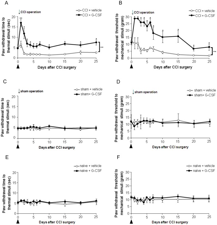
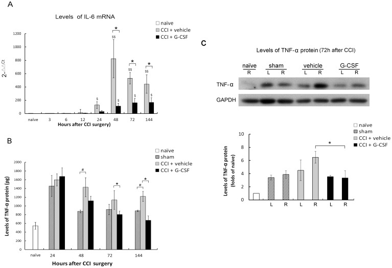
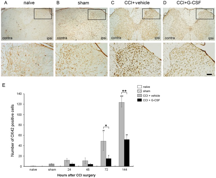
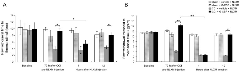
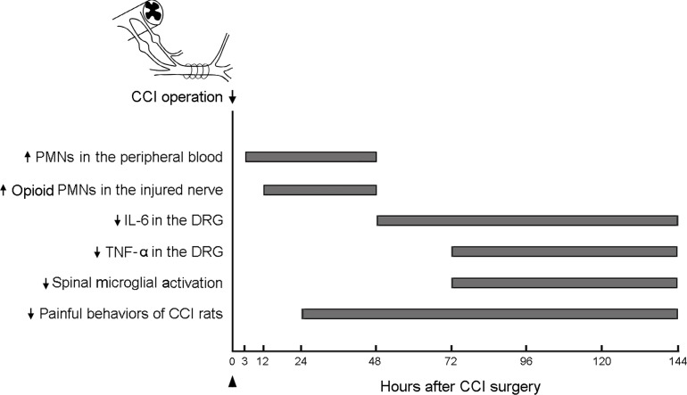
Recent studies have shown that opioid treatment can reduce pro-inflammatory cytokine production and counteract various neuropathic pain syndromes. Granulocyte colony-stimulating factor (G-CSF) can promote immune cell differentiation by increasing leukocytes (mainly opioid-containing polymorphonuclear (PMN) cells), suggesting a potential beneficial role in treating chronic pain. This study shows the effectiveness of exogenous G-CSF treatment (200 µg/kg) for alleviating thermal hyperalgesia and mechanical allodynia in rats with chronic constriction injury (CCI), during post-operative days 1-25, compared to that of vehicle treatment. G-CSF also increases the recruitment of opioid-containing PMN cells into the injured nerve. After CCI, single administration of G-CSF on days 0, 1, and 2, but not on day 3, relieved thermal hyperalgesia, which indicated that its effect on neuropathic pain had a therapeutic window of 0-48 h after nerve injury. CCI led to an increase in the levels of interleukin-6 (IL-6) mRNA and tumor necrosis factor-α (TNF-α) protein in the dorsal root ganglia (DRG). These high levels of IL-6 mRNA and TNF-α were suppressed by a single administration of G-CSF 48-144 h and 72-144 h after CCI, respectively. Furthermore, G-CSF administered 72-144 h after CCI suppressed the CCI-induced upregulation of microglial activation in the ipsilateral spinal dorsal horn, which is essential for sensing neuropathic pain. Moreover, the opioid receptor antagonist naloxone methiodide (NLXM) reversed G-CSF-induced antinociception 3 days after CCI, suggesting that G-CSF alleviates hyperalgesia via opioid/opioid receptor interactions. These results suggest that an early single systemic injection of G-CSF alleviates neuropathic pain via activation of PMN cell-derived endogenous opioid secretion to activate opioid receptors in the injured nerve, downregulate IL-6 and TNF-α inflammatory cytokines, and attenuate microglial activation in the spinal dorsal horn. This indicates that G-CSF treatment can suppress early inflammation and prevent the subsequent development of neuropathic pain.


Conflict of interest statement

Competing Interests: The authors have declared that no competing interests exist.

Figures

Figure 1
Figure 1. G-CSF alleviates long-term thermal hyperalgesia and mechanical allodynia in rats with CCI.
(A) Thermal hyperalgesia was significantly developed in CCI rats treated with vehicle, compared to the pre-operation (p<0.01) and sham-operation (p<0.05) status and persisted throughout the experimental period (25 days). In contrast, G-CSF treatment significantly attenuated the development of thermal hyperalgesia from 1 to 25 days post-CCI compared to the vehicle treatment control. (B) Mechanical allodynia was significantly developed compared to the pre-operation status and persisted throughout the experimental period in CCI rats treated with vehicle; G-CSF treatment alleviated mechanical allodynia from 1 to 25 days post-CCI compared to the vehicle treatment control. (C, D) Compared to vehicle treatment, G-CSF treatment did not alter thermal and mechanical responses in sham-operated rats. (E, F) Similarly, no effect was observed throughout the experimental period on thermal and mechanical responses in naïve control rats treated with G-CSF compared to those treated with vehicle. Data are shown as the means±SEM, n = 6 per group, two-way repeated measures ANOVA, *p<0.05, **p<0.01. Arrows indicate the CCI operation times; arrowheads indicate the time of injection of G-CSF or vehicle.
Figure 2
Figure 2. G-CSF demonstrates a therapeutic time window (0–48 h) after nerve injury.
One-day (A) and two-day (B) delayed injection of G-CSF significantly reversed thermal hyperalgesia compared to the vehicle control, which maintained less thermal hyperalgesia during the entire experiment period. Data are shown as the means±SEM, n = 6 per group, two-way repeated measures ANOVA, *p<0.05. Arrows indicate the CCI operation times; arrowheads indicate the G-CSF or vehicle injection time.
Figure 3
Figure 3. Effects of G-CSF on mobilized immune cells in the tissue fluid around the injured nerves.
Immune cell subpopulations of tissue fluid were quantified by flow cytometry. (A) At 48 h, increased expression of markers, such as, PMN cell marker (stained using anti-PMN-FITC), and β-endorphin marker (stained using anti-β-endorphin-PE), on leukocytes is observed in G-CSF–treated rats with CCI than in vehicle-treated rats with CCI (n = 6 per group). CD45-PE-Cy5 and ED2-PE antibodies were used to react with all hematopoietic cells and macrophages, respectively. Low staining with the respective control antibodies, confirmed a specific antibody staining of opioid-containing PMN cells. In contrast, low staining with ED2-PE and PMN-FITC antibodies (0.3–3.4%), confirmed a specific staining of PMN cells by PMN-FITC antibody. (B) At 48 h, the number of β-endorphin–containing PMN cells in G-CSF–treated rats with CCI is significantly higher than that in vehicle-treated rats with CCI. Data are shown as the mean±SEM, n = 6 per group; t test, *p<0.05.
Figure 4
Figure 4. G-CSF treatment increases the number of migrated β-endorphin-containing PMN cells in injured nerves.
(A–C) Confocal laser scanning microscopy of PMN cells and β-endorphin peptides in the right sciatic nerve of vehicle-treated sham rats (A1–3), vehicle-treated CCI rats (B1–3), and G-CSF treated CCI rats (C1–3) 12 h after CCI, respectively. No apparent PMN cells or β-endorphin peptides were observed in the vehicle-treated sham operation rats. However, at 12 h, the count of β-endorphin-containing PMN cells was significantly higher in the G-CSF-treated CCI rats than in the vehicle-treated CCI rats. Scale bar: 100 µm. (D) Confocal high-power field laser scanning pictures showing a PMN cell with a typical segmented nucleus, which stained positive for β-endorphin peptides. Green: PMN cells; red: β-endorphin (END)-containing cells; blue (DAPI): cell nuclei. Scale bar: 10 µm. (E) The number of β-endorphin-containing PMN cells per section as calculated at different time points. The count of β-endorphin-containing PMN cells was higher in the CCI rats treated with G-CSF (black bars) compared to those treated with vehicle (grey bars) between 12–48 h after nerve injury. Data are shown as the means±SEM, n = 5 per group; one-way ANOVA, *p<0.05, **p<0.01: CCI + G-CSF group compared to CCI + vehicle group; #p<0.05, ##p<0.01: CCI + G-CSF or CCI + vehicle group compared to vehicle-treated sham operation control.
Figure 5
Figure 5. G-CSF reduces the levels of IL-6 mRNA and TNF-α protein in the DRG after CCI.
(A) Quantitative real-time PCR revealed significantly lower levels of IL-6 mRNA in G-CSF-treated CCI rats than in those treated with vehicle 48 and 144 h after nerve injury. Data are shown as the means±SEM, n = 5 per group; one-way ANOVA, *p<0.05: CCI + G-CSF group compared to CCI + vehicle group; $p<0.05, $$p<0.01: CCI + G-CSF or CCI + vehicle group compared to naïve controls. (B) ELISA analysis showed significantly lower levels of TNF-α in G-CSF–treated rats with CCI than in vehicle-treated rats with CCI at 72 and 144 h after nerve injury. Data are shown as the mean±SEM, n = 5 per group; one-way ANOVA, *p<0.05: CCI + G-CSF group compared to CCI + vehicle group; #p<0.05: CCI + G-CSF or CCI + vehicle group compared to vehicle-treated sham-operated controls. (C) Western blot analysis showed significantly lower levels of TNF-α in G-CSF–treated rats with CCI than in vehicle-treated rats with CCI at 72 h after nerve injury. Data are shown as the mean±SEM; n = 5 per group; t test, *p<0.05: CCI + G-CSF group compared to CCI + vehicle group. R: right side; L: left side.
Figure 6
Figure 6. G-CSF reduces immunoreactivity of OX-42 in the spinal cord 72 h after CCI.
Representative images show the OX-42 peptides in the right L5 dorsal horn of (A) naïve, (B) sham operation rats, (C) vehicle-treated CCI rats, and (D) G-CSF-treated CCI rats. There was a basal and mild activation of OX-42 immunoreactivity in naïve and sham operation rats, respectively. A significantly higher level of OX-42 immunoreactivity was observed in CCI rats treated with vehicle. In contrast, a significantly lower level of OX-42 immunoreactivity (similar to the sham-operation rats) was noted 72 h post-CCI in the CCI rats treated with G-CSF than those treated with vehicle. (E) Quantification of OX-42 immunoreactivity in L5 dorsal horn after vehicle- or G-CSF- treated CCI rats 72 h post-CCI. Data are shown as the means±SEM, n = 5 per group; one-way ANOVA, *p<0.05, **p<0.01: CCI + G-CSF group compared to CCI + vehicle group; #p<0.05, ##p<0.01: CCI + G-CSF or CCI + vehicle group compared with naïve or sham operation controls. ipsi: ipsilateral (right) side; contra: contralateral (left) side. Scale bar: 50 µm.
Figure 7
Figure 7. NLXM reverses the G-CSF anti-nociceptive effect.
The histograms represent the effect of opioid/opioid receptor interaction on thermal and mechanical responses in sham or CCI rats with/without G-CSF treatment 72 h post-CCI operation, followed by a local injection of NLXM. One hour after injection, NLXM reversed G-CSF antinociception effect on both thermal hyperalgesia (A) and mechanical allodynia (B); however, the effect of NLXM lasted for 1 h but did not show any further reversal effects on antinociception at 12 h post-injection. In contrast, NLXM did not alter the thermal and mechanical responses in sham-operated rats with/without G-CSF treatment. Data are shown as the means±SEM, n = 6 per group; two-way repeated measures ANOVA, *p<0.05, **p<0.01: CCI + G-CSF group compared to CCI + vehicle group; #p<0.05, ##p<0.01: CCI + G-CSF group compared to CCI + G-CSF + NLXM.
Figure 8
Figure 8. The time sequence of the effects of G-CSF on pain, cytokine levels, and microglial activation.
The sequential effect of G-CSF in rats with CCI are po (post-operative) increase in PMN cells and opioid-containing PMN cells in the peripheral blood at 3 h and in the injured nerves at 12 h, downregulation of the levels of IL-6 within po 48–144 h and TNF-α within po 72–144 h in the DRG, and suppression of spinal microglial activation within po 72–144 h, all of which are associated with long-term pain alleviation after nerve injury. The arrow indicates the time at which the operation for CCI was performed; the arrowhead indicates the G-CSF injection time.

References

    1. Marchand F, Perretti M, McMahon SB (2005) Role of the immune system in chronic pain. Nat Rev Neurosci 6: 521–532. - PubMed
    1. Fecho K, Manning EL, Maixner W, Schmitt CP (2007) Effects of carrageenan and morphine on acute inflammation and pain in Lewis and Fischer rats. Brain Behav Immun 21: 68–78. - PubMed
    1. Finley MJ, Happel CM, Kaminsky DE, Rogers TJ (2008) Opioid and nociceptin receptors regulate cytokine and cytokine receptor expression. Cell Immunol 252: 146–154. - PMC - PubMed
    1. Berrios I, Castro C, Kuffler DP (2008) Morphine: axon regeneration, neuroprotection, neurotoxicity, tolerance, and neuropathic pain. P R Health Sci J 27: 119–128. - PubMed
    1. Ugolini G, Marinelli S, Covaceuszach S, Cattaneo A, Pavone F (2007) The function neutralizing anti-TrkA antibody MNAC13 reduces inflammatory and neuropathic pain. Proc Natl Acad Sci U S A 104: 2985–2990. - PMC - PubMed

Publication types

MeSH terms

Substances