. 2012;7(8):e43733.
doi: 10.1371/journal.pone.0043733. Epub 2012 Aug 24.

Mtb-specific CD27low CD4 T cells as markers of lung tissue destruction during pulmonary tuberculosis in humans

Affiliations

Mtb-specific CD27low CD4 T cells as markers of lung tissue destruction during pulmonary tuberculosis in humans

Irina Yu Nikitina et al. PLoS One. 2012.

Abstract

Background: Effector CD4 T cells represent a key component of the host's anti-tuberculosis immune defense. Successful differentiation and functioning of effector lymphocytes protects the host against severe M. tuberculosis (Mtb) infection. On the other hand, effector T cell differentiation depends on disease severity/activity, as T cell responses are driven by antigenic and inflammatory stimuli released during infection. Thus, tuberculosis (TB) progression and the degree of effector CD4 T cell differentiation are interrelated, but the relationships are complex and not well understood. We have analyzed an association between the degree of Mtb-specific CD4 T cell differentiation and severity/activity of pulmonary TB infection.

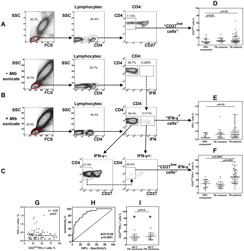
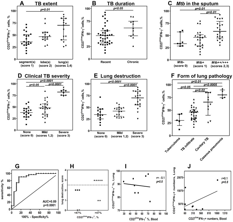
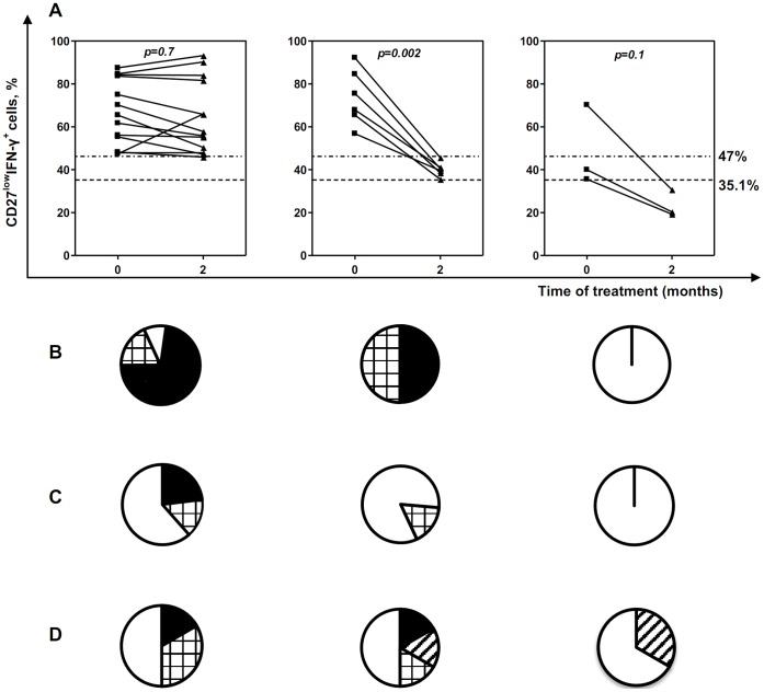
Methodology/principal findings: The degree of CD4 T cell differentiation was assessed by measuring the percentages of highly differentiated CD27(low) cells within a population of Mtb- specific CD4 T lymphocytes ("CD27(low)IFN-γ(+)" cells). The percentages of CD27(low)IFN-γ+ cells were low in healthy donors (median, 33.1%) and TB contacts (21.8%) but increased in TB patients (47.3%, p<0.0005). Within the group of patients, the percentages of CD27(low)IFN-γ(+) cells were uniformly high in the lungs (>76%), but varied in blood (12-92%). The major correlate for the accumulation of CD27(low)IFN-γ(+) cells in blood was lung destruction (r = 0.65, p = 2.7 × 10(-7)). A cutoff of 47% of CD27(low)IFN-γ(+) cells discriminated patients with high and low degree of lung destruction (sensitivity 89%, specificity 74%); a decline in CD27(low)IFN-γ(+)cells following TB therapy correlated with repair and/or reduction of lung destruction (p<0.01).

Conclusions: Highly differentiated CD27(low) Mtb-specific (CD27(low)IFN-γ(+)) CD4 T cells accumulate in the lungs and circulate in the blood of patients with active pulmonary TB. Accumulation of CD27(low)IFN-γ(+) cells in the blood is associated with lung destruction. The findings indicate that there is no deficiency in CD4 T cell differentiation during TB; evaluation of CD27(low)IFN-γ(+) cells provides a valuable means to assess TB activity, lung destruction, and tissue repair following TB therapy.


Conflict of interest statement

Competing Interests: The authors have declared that no competing interests exist.

Figures

Figure 1
Figure 1. Evaluation of lung tissue destruction.
Examples of X-ray computer tomograpy with different degrees of tissue destruction are shown. Lung destruction was evaluated based on the number and size of destructive (lucent) foci (see Methods for the details). A, score 0, no destruction; B, score 1, one small (<2 cm) destruction; C, score 2, one large (>2 cm) destruction; D, score 3, multiple lung destructions. Arrows, lung destructions. Asterisk, infiltrative focus without destruction.
Figure 2
Figure 2. TB patients have increased percentages of CD27lowIFN-γ+ CD4 T cells in their blood.
A–C, Strategies for determining percentages of CD27low (A), IFN-γ+ (B) and CD27lowIFN-γ+ (C) CD4 T cells. A, CD27low cells were gated within the total population of CD4+ T cells. B, To identify IFN-γ+ CD4 T cells, an aliquot of blood was stimulated with Mtb sonicate; another aliquote was left un-stimulated. During the analysis, the gates for IFN-γ+ cells in Mtb-stimulated samples were plotted based on Mtb un-stimulated samples (Fig. B, dotted line). To identify CD27lowIFN-γ+ cells, the expression of CD27 was first analyzed in IFN-γ population. Because this population was always numerous, CD27low and CD27hi cells could be easily separated. The gates for CD27low cells were then applied to IFN-γ+ population (C, dotted line). D–F, Percentages of CD27low (D), IFN-γ+ (E), and CD27lowIFN-γ+ (F) cells in TB patients (n = 50), TB contacts (n = 21) and Mtb-unexposed individuals (n = 15). G, Lack of correlation between the percentages of IFN-γ+ and CD27lowIFN-γ+ cells in TB patients, TB contacts and Mtb-unexposed individuals (n = 86). H, ROC-curve of CD27lowIFN- γ + cell percentages for discriminating TB patients from healthy individuals (TB contacts and Mtb-unexposed). I, Percentages of CD27lowIFN-γ+ cells in TB contacts with positive and negative results of QFT assay *p<0.0005 compared to TB patients.
Figure 3
Figure 3. Association between blood CD27lowIFN-γ+ cells and different manifestations of TB disease.
A–F, Percentages of CD27lowIFN-γ+ cells in TB patients (n = 50) grouped based on different characteristics of TB disease. For multiple (seven) parameter testing, p- value <0.007 was considered significant. G, ROC curve for discriminating TB patients with high (score 3) and low (scores 0–2) degrees of lung tissue destruction (n = 50). H, The degree of lung tissue destruction in patients with “low” (<47%) and high (>47%) percentages of CD27lowIFN-γ+ CD4 T cells (n = 12, validation analysis). I, J, Lack of correlation between the percentages (I) and the numbers (J) of CD27lowIFN-γ+ cells in the lungs and in the blood of TB patients (n = 8). Indicated are numbers of CD27lowIFN-γ+ cells per 1 million of acquired cells.
Figure 4
Figure 4. Following anti-TB therapy, CD27lowIFN-γ+ cells decline parallel to reduction/repair of lung destruction.
A, Percentages of CD27lowIFN-γ+ cells were determined at the start of treatment and two months later. Depending on the results of “IFN-γ/CD27” assay, patients were divided into three groups: in “CD27low- high” group, initially elevated percentages of CD27lowIFN-γ+ cells did not decline following 2-mo therapy; in “CD27low – reduced” group, percentages of CD27lowIFN-γ+ cells declined to become below the 47% threshold, but remained above 35.1% threshold (upper limit of norm); in “CD27low - normalized” group, percentages of CD27lowIFN-γ+ cells declined to become below the 35.1% threshold. Dotted lines show 35.1 and 47% thresholds. B–D, diagrams showing changes in lung destruction (B), sputum Mtb-positivity (C) and clinical TB severity (D) for each group of patients following 2-mo therapy. Closed segments, no improvement; squared segments, reduction of lung destruction, clinical symptoms or numbers of Mtb in the sputum; open segments, normalization (repair of lung destruction, conversion of sputum assay, disappearance of intoxication symptoms), striped segments – no abnormalities at the start of the treatment.

References

    1. Jasmer RM, Nahid P, Hopewell PC (2002) Clinical practice. Latent tuberculosis infection. N Engl J Med 347: 1860–1866. - PubMed
    1. Wallis RS, Pai M, Menzies D, Doherty TM, Walzl G, et al. (2010) Biomarkers and diagnostics for tuberculosis: progress, needs, and translation into practice. Lancet 375: 1920–1937. - PubMed
    1. den Hertog AL, Mayboroda OA, Klatser PR, Anthony RM (2011) Simple Rapid Near-Patient Diagnostics for Tuberculosis Remain Elusive-Is a “Treat-to-Test” Strategy More Realistic? PLoS Pathol 7. Available: http://www.plospathogens.org/article/info:doi/10.1371/journal.ppat.10022... via the Internet. Accessed 2011 Nov 3. - DOI - PMC - PubMed
    1. Ewer K, Deeks J, Alvarez L, Bryant G, Waller S, et al. (2003) Comparison of T- cell-based assay with tuberculin skin test for diagnosis of Mycobacterium tuberculosis infection in a school tuberculosis outbreak. Lancet 361: 1168–1173. - PubMed
    1. Mori T, Sakatani M, Yamagishi F, Takashima T, Kawabe Y, et al. (2004) Specific detection of tuberculosis infection: an interferon-gamma-based assay using new antigens. Am J Respir Crit Care Med 170: 59–64. - PubMed

Publication types

Substances