. 2012 Aug;5(4):230-7.
doi: 10.1593/tlo.12136. Epub 2012 Aug 1.

An integrated method for reproducible and accurate image-guided stereotactic cranial irradiation of brain tumors using the small animal radiation research platform

Affiliations

An integrated method for reproducible and accurate image-guided stereotactic cranial irradiation of brain tumors using the small animal radiation research platform

Brian C Baumann et al. Transl Oncol. 2012 Aug.

Abstract

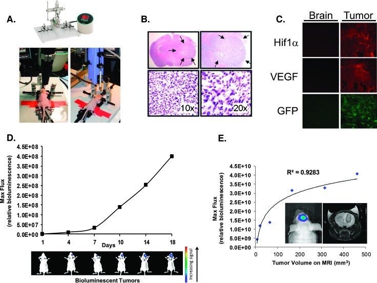
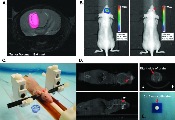
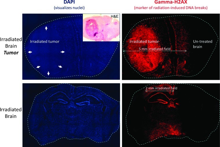
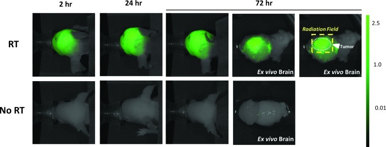
Preclinical studies of cranial radiation therapy (RT) using animal brain tumor models have been hampered by technical limitations in the delivery of clinically relevant RT. We established a bioimageable mouse model of glioblastoma multiforme (GBM) and an image-guided radiation delivery system that facilitated precise tumor localization and treatment and which closely resembled clinical RT. Our novel radiation system makes use of magnetic resonance imaging (MRI) and bioluminescent imaging (BLI) to define tumor volumes, computed tomographic (CT) imaging for accurate treatment planning, a novel mouse immobilization system, and precise treatments delivered with the Small Animal Radiation Research Platform. We demonstrated that, in vivo, BLI correlated well with MRI for defining tumor volumes. Our novel restraint system enhanced setup reproducibility and precision, was atraumatic, and minimized artifacts on CT imaging used for treatment planning. We confirmed precise radiation delivery through immunofluorescent analysis of the phosphorylation of histone H2AX in irradiated brains and brain tumors. Assays with an intravenous near-infrared fluorescent probe confirmed that radiation of orthografts increased disruption of the tumor blood-brain barrier (BBB). This integrated model system, which facilitated delivery of precise, reproducible, stereotactic cranial RT in mice and confirmed RT's resultant histologic and BBB changes, may aid future brain tumor research.


Figures

Figure 1
Figure 1
Establishing human-derived orthotopic GBM tumors in mice and subsequent imaging with bioluminescent and MR imaging. (A) Stereotactic implantation of GBM cells expressing GFP and luciferase into the brains of nude mice to create orthotopic tumors. (B) Hematoxylin/eosin staining of brains excised from mice shows the orthotopic tumor, designated within the arrows, under light microscopy at four different magnifications. (C) Orthotopic GBM tumors show robust expression of HIF-1α and VEGF. Normal brain (left column) or orthotopic brain tumor tissue (right column) were sectioned, fixed onto slides, and stained for HIF-1α (top) or VEGF (middle) protein expression. The bottom row shows expression of GFP. The immunofluorescent analyses were performed through the respective specific primary antibodies followed by red fluorescent protein (RFP)-labeled secondary antibodies to detect the specific signal. (D) BLI shows serial growth of the implanted tumors (representative mice shown during BLI on the specified day after initial implantation of tumor cells). Bioluminescence flux also reflects intracranial growth patterns. GBM cells expressing GFP and luciferase were stereotactically implanted, followed by in vivo serial BLI of the resultant brain tumors. BLI flux was defined as photons per second per squared centimeter, which reliably reflected intracranial growth patterns. (E) Bioluminescent imaging signal from tumors growing within the mouse correlate well with tumor volume measured after excision. In vivo BLI signal (“Bioluminescent Max Flux”) was recorded for mice orthotopic brain tumors immediately before sacrifice. The tumors of each mouse were then excised, and the respective volume of each tumor was manually measured. The measured tumor volumes significantly correlate with BLI values (R2 = 0.9283).
Figure 2
Figure 2
Image-guided stereotactic delivery of radiation to implanted tumors. (A) Spin-echo MRI of a mouse intracranial tumor using a 9.4-T MR spectrometer to determine the precise anatomic localization of the xenograft. The tumor volume was contoured in pink using the software package Amira. (B) Point of maximal bioluminescent signal for the tumor was determined (red arrow), and the spot was tattooed to aid in the setup of radiation treatment. (C) Anesthetized mouse secured in the home-built novel stereotactic restrainer. The point of maximal bioluminescent signal, identified previously, is covered by a fiducial marker (red arrow) to facilitate identification of the location on CT imaging for treatment planning. (D) Treatment planning CT scans showing the coronal, axial, and sagittal views of the brain. The small red crosses in each image indicate the selected isocenter. In the top right image, the coronal CT of the brain reveals three coplanar fiducial markers used to confirm proper setup for treatment (the scalp fiducial marker and two lateral fiducial markers on the restrainer seen at the bottom corners). (E) Irradiation of exposure film using the 5 x 5-mm collimator confirmed that the SARRP can deliver a collimated beam of radiation with very limited scatter. The irradiated field was centered within ±0.25 mm on its isocenter.
Figure 3
Figure 3
γH2AX staining of irradiated brain tissue confirms accurate delivery of stereotactic brain irradiation. Top row shows fluorescent microscopy images of coronal brain sections from a mouse with an intracranial (forebrain) tumor (marked with arrows) killed 2 hours after focal RT to 20 Gy in a single fraction using a 5 x 5-mm collimator, followed by staining for DAPI to visualize cell nuclei (left) and γH2AX to visualize radiation-induced unrepaired double-strand breaks (right). γH2AX staining confirms the precise targeting of the tumor using the described technique. The bottom row shows DAPI and γH2AX staining performed on coronal midbrain sections of a mouse without intracranial tumor killed 2 hours after focal RT to 20 Gy x 1 using a 7 x 2-mm collimator with the long axis parallel to the animal's spine.
Figure 4
Figure 4
Targeted cranial RT for GBM orthografts is associated with increased extravasation of a near-infrared pegylated fluorescent probe, thus indicating disruption of the tumor BBB. Mice with intracranial tumors of comparable sizes (confirmed by tumor BLI signal) were irradiated through the SARRP (“RT,” 12 Gy in four daily fractions) or mock irradiated (“No RT”). After completion of RT, mice were injected through the tail vein with LI-COR pegylated Near IR Dye. Serial in vivo fluorescent imaging revealed significantly increased fluorescent signal in the irradiated brain tumors at all time points, indicating increased BBB disruption induced by the focused radiation delivered. The group of three panels on the right (each labeled “Brain”) shows the tumor-containing irradiated (top) or mock-irradiated (bottom) brains imaged after removal from the mouse (“ex vivo imaging”). The far right image confirms that increased BBB disruption within the brain tumor corresponded to the RT field. The dashed box delineates the RT field, within which extravasation of near-infrared fluorescent probe was maximal.

References

    1. Stupp R, Mason WP, van den Bent MJ, Weller M, Fisher B, Taphoorn MJ, Belanger K, Brandes AA, Marosi C, Bogdahn U, et al. Radiotherapy plus concomitant and adjuvant temozolomide for glioblastoma. N Engl J Med. 2005;352:987–996. - PubMed
    1. Zhou H, Rodriguez M, van den Haak F, Nelson G, Jogani R, Xu J, Zhu X, Xian Y, Tran PT, Felsher DW, et al. Development of a micro-computed tomography-based image-guided conformal radiotherapy system for small animals. Int J Radiat Oncol Biol Phys. 2010;78:297–305. - PMC - PubMed
    1. Wong J, Armour E, Kazanzides P, Iordachita I, Tryggestad E, Deng H, Matinfar M, Kennedy C, Liu Z, Chan T, et al. High-resolution, small animal radiation research platform with x-ray tomographic guidance capabilities. Int J Radiat Oncol Biol Phys. 2008;71:1591–1599. - PMC - PubMed
    1. Stojadinovic S, Low DA, Hope AJ, Vicic M, Deasy JO, Cui J, Khullar D, Parikh PJ, Malinowski KT, Izaguirre EW, et al. MicroRT—small animal conformal irradiator. Med Phys. 2007;34:4706–4716. - PubMed
    1. Baumann BC, Dorsey JF, Benci JL, Joh DY, Kao GD. Stereotactic intracranial implantation and in vivo bioluminescent imaging of tumor xenografts in a mouse model system of glioblastoma multiforme. J Vis Exp. 2012 In press. - PMC - PubMed

LinkOut - more resources