. 2012 Aug;5(4):260-8.
doi: 10.1593/tlo.12184. Epub 2012 Aug 1.

Expression of death receptor 6 by ovarian tumors in laying hens, a preclinical model of spontaneous ovarian cancer

Affiliations

Expression of death receptor 6 by ovarian tumors in laying hens, a preclinical model of spontaneous ovarian cancer

Animesh Barua et al. Transl Oncol. 2012 Aug.

Abstract

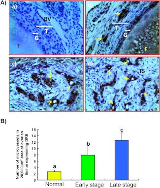
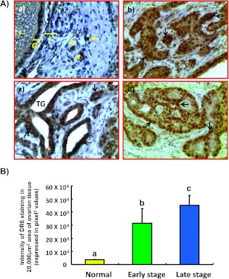
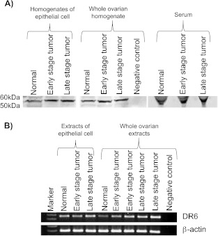
Tumor-associated neoangiogenesis and suppression of antitumor immunity are hallmarks of tumor development and progression. Death receptor 6 (DR6) has been reported to be associated with suppression of antitumor immunity and tumor progression in several malignancies. However, expression of DR6 by malignant ovarian epithelial tumors at an early stage is unknown. The goals of this study were to determine whether DR6 is expressed by malignant ovarian epithelial tumors at an early stage and to examine whether DR6 expression is associated with ovarian cancer (OVCA) progression in a laying hen model of spontaneous OVCA. Expression of DR6 was examined in normal and malignant ovaries, normal ovarian surface epithelial (OSE) cells, or malignant epithelial cells and in serum of 3-year-old hens. The population of microvessels expressing DR6 was significantly higher in hens with early-stage OVCA than hens with normal ovaries (P < .01) and increased further in late-stage OVCA. The results of this study showed that, in addition to microvessels, tumor cells in the ovary also express DR6 with a significantly higher intensity than normal OSE cells. Similar patterns of DR6 expression were also observed by immunoblot analysis and gene expression studies. Furthermore, DR6 was also detected in the serum of hens. In conclusion, DR6 expression is associated with OVCA development and progression in laying hens. This study may be helpful to examine the feasibility of DR6 as a useful surrogate marker of OVCA, a target for antitumor immunotherapy and molecular imaging and thus provide a foundation for clinical studies.


Figures

Figure 1
Figure 1
Gross morphology of normal and malignant ovaries in laying hens. (A) Fully functional normal ovary in a laying hen. Laying hens ovulate and subsequently lay eggs once a day for 5 or 6 days in a week in a continuous manner and then take a pause for 1 day before laying resumes. Thus, the ovary in a fully functional laying hen contains a set of multiple large and growing preovulatory follicles arranged in a hierarchy of sizes (F1–F5). The largest follicle (F1) is destined to ovulate soon and then the second largest follicle (F2) becomes F1 and a small growing follicle is recruited from the ovarian stroma to the hierarchy to maintain the laying rates. (B) Ovarian tumor at an early stage in a hen showing solid tumor mass (arrows) limited to the ovary. (C) Ovarian tumor at a late stage. The2 tumor (Tu) appears like a cauliflower and has metastasized to other organs with accompanied profuse ascites (*).
Figure 2
Figure 2
Microscopic features of hen ovaries. Paraffin-embedded sections from normal or malignant ovaries with tumors were stained with hematoxylin and eosin. (A) Section of a normal ovary showing a developing follicle embedded in the ovarian stroma. (B) Section of an ovarian serous carcinoma showing a solid sheet of tumor surrounded by fibromuscular tissue. The tumor contains a labyrinth of slitlike glandular spaces lined by cells with large pleomorphic nuclei and mitotic figures. (C) Section showing endometrioid carcinoma displaying confluent back-to-back glands. Glands contain a single layer of epithelial cells with sharp luminal margins. (D) Section of a mucinous carcinoma. Glands in clusters with scarce intervening stroma lined by columnar and goblet cells with intracytoplasmic mucin. Original magnification, x40.
Figure 3
Figure 3
(A) Immunohistochemical detection of DR6-expressing microvessels in hen ovaries with or without tumor. (a) Section of a normal ovarian stroma immunostained by omitting primary antibodies used as control. No immunopositive vessel is seen. (b) Serial section from the same normal ovary immunostained with primary antibodies. Very few DR6-expressing vessels are seen. (c) An ovarian section from a hen with early-stage OVCA. Compared to the normal ovary, many DR6-expressing microvessels are seen in the stroma between tumors. (d) Section of a malignant tumor from a hen with late-stage OVCA. Many DR6-expressing microvessels are localized in the tumor stroma. BV indicates blood vessel; F, follicle; G, granulosa layer; T, theca layer; TS, tumor stroma. Arrows indicate DR6-expressing microvessels. Original magnification, x40. (B) Changes in the frequency of DR6-expressing ovarian microvessels relative to ovarian tumor development and progression in hens. The frequency of microvessels expressing DR6 in a 20,000-µm2 area of normal (n = 15) and malignant ovaries (expressed as the mean ± SD, n = 15 each for early and late stages). Compared to the normal ovary, the frequency of microvessels expressing DR6 was significantly (P < .001) higher in hens with early-stage OVCA cancer and increased further (P < .001) as the disease progressed to a late stage in hens. Each bar with a different letter indicates significant differences (P < .001) between normal and tumor groups.
Figure 4
Figure 4
(A) Expression of DR6 by tumor cells in malignant ovaries in hens. (a) Section of a normal ovary immunostained for DR6 expression. Very few immunopositive cells are present in the ovarian stroma. (b-d) Sections of different histologic subtypes of OVCA in hens including serous (b), endometrioid (c), and mucinous (d) at early stages immunostained for DR6 expression. Compared with the normal OSE cells, tumor cells in all three histologic subtypes stained intensely. Original magnification, x40. (B) Changes in the DR6 staining intensity relative to ovarian tumor development and progression in hens. The intensity of DR6 staining is expressed as the pixel values (mean ± SD) in a 20,000-µm2 area in normal ovarian stroma (n = 15) or in malignant ovaries. Compared to the normal ovary, the intensity of DR6 staining was significantly (P < .001) higher in hens with early-stage OVCA (n = 15) and increased further (P < .001) in late-stage OVCA (n = 15). Each bar with different letter indicates significant differences (P < .001) among normal or OVCA stages.
Figure 5
Figure 5
Immunoreactive (A) DR6 protein or (B) mRNA in serum and ovaries of hens with normal ovaries or those with OVCA. (A) One-dimensional Western blot analysis: Immunoreactive DR6 proteins of 50-to 60-kDa molecular weight were detected in the homogenates of normal OSE cells or tumor cells, in homogenates of whole normal or malignant ovaries, as well as in serum of hens by one-dimensional Western blot. Compared to the hens with normal ovaries, relatively stronger immunoreactive bands for DR6 proteins were observed in serum and ovaries of hens with early and late stages of OVCA. No immunoreactive band was detected in the negative control in which protein sample was omitted. (B) Semiquantitative RT-PCR: mRNA expression for DR6 was detected in the extracts of normal OSE cells or tumor cells and in extracts of normal and malignant ovaries by semiquantitative PCR. Compared to the weak expression by normal ovaries and OSE, strong amplification for DR6 mRNA was observed in the extracts of malignant ovaries and tumor cells in hens with early- and late-stage OVCA. No DR6 mRNA expression was detected in the negative control in which mRNA sample was omitted.

References

    1. Jemal A, Siegel R, Xu J, Ward E. Cancer statistics, 2010. CA Cancer J Clin. 2010;60:277–300. - PubMed
    1. Ferlay J, Autier P, Boniol M, Heanue M, Colombet M, Boyle P. Estimates of the cancer incidence and mortality in Europe in 2006. Ann Oncol. 2007;18:581–592. - PubMed
    1. Liu VC, Wong LY, Jang T, Shah AH, Park I, Yang X, Zhang Q, Lonning S, Teicher BA, Lee C. Tumor evasion of the immune system by converting CD4+CD25- T cells into CD4+CD25+ T regulatory cells: role of tumor-derived TGF-β. J Immunol. 2007;178:2883–2892. - PubMed
    1. Curiel TJ. Tregs and rethinking cancer immunotherapy. J Clin Invest. 2007;117:1167–1174. - PMC - PubMed
    1. Yigit R, Massuger LF, Figdor CG, Torensma R. Ovarian cancer creates a suppressive microenvironment to escape immune elimination. Gynecol Oncol. 2010;117:366–372. - PubMed

LinkOut - more resources